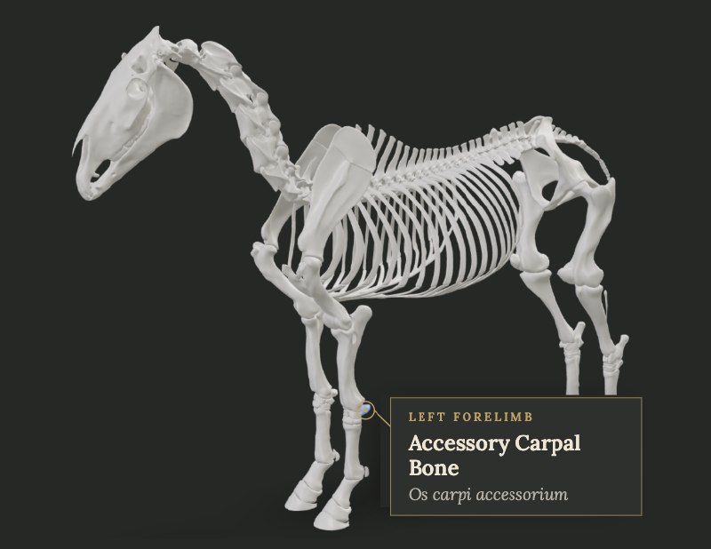
Accessory Carpal Bone (Left)
Os carpi accessorium (Ca)
Overview
The accessory carpal bone (os carpi accessorium, Ca) is a distinctive, non-weight-bearing element of the proximal row of the equine carpus. It projects palmarolaterally from the carpus, articulating primarily with the ulnar carpal bone and the caudal aspect of the distal radius (including the lateral styloid process region). Its medial surface is concave and fibrocartilage-covered, forming the lateral wall of the carpal canal (carpal sheath), through which the superficial and deep digital flexor tendons, the carpal check ligament, and associated vessels and nerves pass. Laterally it presents a groove that accommodates the tendon of insertion of ulnaris lateralis (extensor carpi ulnaris). Four major ligaments stabilize it: accessorioulnar, accessoriocarpoulnar, accessorioquartal, and accessoriometacarpal. Functionally it serves as a lever arm that increases the mechanical advantage of the flexor carpi ulnaris and ulnaris lateralis, contributing to carpal flexion and shock absorption during loading. Because it is largely non-articular on its palmar and lateral faces and is subjected to strong tensile forces from the flexor musculature, it is vulnerable to fracture under hyperextension, direct trauma, or asynchronous muscle pull. The bone is readily palpable on the lateral-palmar aspect of the "knee." Blood supply is via branches of the median and radial arteries; innervation is sensory via branches of the median and ulnar nerves. In the standing horse it helps maintain the integrity of the carpal canal and the position of the flexor tendons. During locomotion its motion is not purely passive; recent kinematic studies show it moves independently relative to the radius and third metacarpus, especially in flexion. This mobility, combined with the constant tension of attached muscles and ligaments, explains why fractures often heal by fibrous union rather than solid bony union and why secondary carpal-canal syndrome can develop.
Clinical Significance
Fractures are most often frontal-plane through the midbody (groove of ulnaris lateralis) or avulsions of the dorsoproximal or palmarodistal margins. Mechanisms include direct trauma, hyperextension with "nutcracker" compression between radius and third metacarpus, or asynchronous pull of flexor carpi ulnaris versus ulnaris lateralis. Clinical signs: acute lameness, pain on flexion, swelling of the carpal canal, sometimes reduced digital pulses when the carpus is flexed. Diagnosis: lateromedial and flexed lateromedial radiographs; ultrasound of the carpal sheath to detect tendon laceration or fragment displacement; CT or MRI for complex cases. Treatment: conservative (immobilization) for nondisplaced fractures often results in fibrous union; displaced fractures that communicate with the sheath may require arthroscopic/tenoscopic debridement of fragments and damaged DDFT (deep digital flexor tendon), or lag-screw fixation (technically demanding because of the curved shape). Carpal canal syndrome secondary to effusion or fibrosis is managed by sheath decompression or annular ligament release. Prognosis is fair to good for return to work if secondary soft-tissue damage is limited.
Common Conditions & Injuries
Fractures (from direct trauma or hyperextension)
## Overview Fractures caused by direct trauma or hyperextension occur when a bone is struck or forced beyond its normal range. Common examples include carpal chip fractures from hyperextension in rac
Carpal canal syndrome
## Overview Carpal canal syndrome is compression of structures within the carpal canal (carpal sheath) producing pain and lameness. The canal contains the superficial and deep digital flexor tendons,
What Horse Owners Should Know
Feel the back-outside of each knee for heat, swelling, or pain on light pressure. A horse that suddenly becomes lame and holds the knee slightly flexed, especially after a fall or jump, may have injured this bone. Avoid forcing full flexion if the horse is painful. Call your veterinarian promptly for any acute swelling or lameness behind the knee; early radiographs and ultrasound of the carpal canal improve outcome. Protect the area during trailering and when jumping on hard ground.
Frequently Asked Questions
What is Fractures (from direct trauma or hyperextension) in horses?
## Overview Fractures caused by direct trauma or hyperextension occur when a bone is struck or forced beyond its normal range. Common examples include carpal chip fractures from hyperextension in racehorses and traumatic fractures from kicks or falls. Treatment depends on location and articular involvement. ## Causes Acute overload or impact. ## Diagnosis Radiographs. Arthroscopy for articular fragments. ## Treatment Arthroscopic removal for many chips; internal fixation for larger or unstable fractures. ## Recovery and Prognosis Good for many articular chips. Larger traumatic fractures have a more variable outlook. ## Prevention Conditioning and safe environments. Signs include: acute lameness after trauma or fast work; joint effusion; radiographic fracture
What is Carpal canal syndrome in horses?
## Overview Carpal canal syndrome is compression of structures within the carpal canal (carpal sheath) producing pain and lameness. The canal contains the superficial and deep digital flexor tendons, their accessory ligaments, and the median artery and nerve. Effusion, desmopathy of the accessory ligament of the superficial digital flexor tendon, distal radial osteochondromas, or retinacular thickening can all produce the syndrome. ## Causes Desmopathy of the proximal check ligament is one possible cause. Other causes include radial exostoses, idiopathic tenosynovitis, and trauma. Chronic effusion leads to pressure on the contained soft tissues. ## Diagnosis Forelimb lameness abolished by proximal metacarpal or antebrachial nerve blocks, combined with carpal sheath effusion, is suggestive. Ultrasound is essential to identify the specific lesion within or adjacent to the canal. ## Treatment Rest and controlled exercise for 3 to 6 months, carpal sheath injection, and corrective shoeing are first-line measures. Surgical release or removal of space-occupying lesions is considered when medical management fails. ## Recovery and Prognosis Prognosis is fair to good for many soft-tissue lesions with appropriate rest. Space-occupying bony lesions often require surgery for lasting improvement. ## Prevention No specific prevention is established. Early attention to check-ligament injuries may limit secondary canal compression. Signs include: forelimb lameness; swelling of the carpal sheath; pain on carpal flexion; positive proximal limb nerve blocks
What are the clinical concerns for the Accessory Carpal Bone (Left)?
Fractures are most often frontal-plane through the midbody (groove of ulnaris lateralis) or avulsions of the dorsoproximal or palmarodistal margins. Mechanisms include direct trauma, hyperextension with "nutcracker" compression between radius and third metacarpus, or asynchronous pull of flexor carpi ulnaris versus ulnaris lateralis. Clinical signs: acute lameness, pain on flexion, swelling of the carpal canal, sometimes reduced digital pulses when the carpus is flexed. Diagnosis: lateromedial and flexed lateromedial radiographs; ultrasound of the carpal sheath to detect tendon laceration or fragment displacement; CT or MRI for complex cases. Treatment: conservative (immobilization) for nondisplaced fractures often results in fibrous union; displaced fractures that communicate with the sheath may require arthroscopic/tenoscopic debridement of fragments and damaged DDFT (deep digital flexor tendon), or lag-screw fixation (technically demanding because of the curved shape). Carpal canal syndrome secondary to effusion or fibrosis is managed by sheath decompression or annular ligament release. Prognosis is fair to good for return to work if secondary soft-tissue damage is limited.
What should horse owners know about the Accessory Carpal Bone (Left)?
Feel the back-outside of each knee for heat, swelling, or pain on light pressure. A horse that suddenly becomes lame and holds the knee slightly flexed, especially after a fall or jump, may have injured this bone. Avoid forcing full flexion if the horse is painful. Call your veterinarian promptly for any acute swelling or lameness behind the knee; early radiographs and ultrasound of the carpal canal improve outcome. Protect the area during trailering and when jumping on hard ground.
Explore This Structure in 3D
See the Accessory Carpal Bone (Left) on our interactive 3D horse model. Open 3D Model