Arterial System (Detailed)
Arteriae
Overview
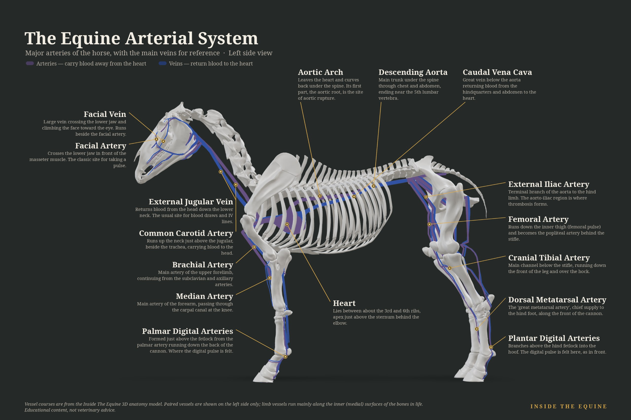
The equine arterial system begins at the left ventricle and is organized around the aorta. The ascending aorta gives rise to the coronary arteries; the aortic arch continues as the brachiocephalic trunk, which supplies the head, neck, and both forelimbs via the common carotid and subclavian arteries and their branches. The descending aorta travels caudally through the thorax and abdomen, giving off intercostal, lumbar, and visceral branches before bifurcating into the external and internal iliac arteries that supply the hind limbs and pelvis. Within the limbs the major arteries (median, radial, femoral, popliteal, etc.) progressively divide until they reach the digit. The medial and lateral digital arteries, running on either side of the deep digital flexor tendon within the hoof, are the final conduits of blood to the sensitive laminae. Any sustained reduction in digital arterial flow or increase in venous resistance can produce laminar ischemia: the central vascular event in many forms of laminitis. Clinically, the strength and character of the digital pulse (palpated over the digital arteries at the fetlock or pastern) is a daily screening tool for inflammation or laminitis. Doppler ultrasonography allows non-invasive assessment of flow velocity and waveform in larger vessels. Pathologic conditions include aortic rupture (often at the root or sinus of Valsalva in older horses or breeding stallions), aorto-iliac thrombosis (exercise-induced hind-limb weakness), verminous arteritis from migrating Strongylus vulgaris larvae (now less common with modern deworming), and peripheral arterial thrombosis or embolism. The arterial walls contain elastic and muscular components that dampen the pulse pressure; age-related or inflammatory changes can stiffen these walls and alter distal perfusion. Understanding the arterial tree is therefore essential for interpreting lameness, colic of vascular origin, and performance problems linked to inadequate oxygen delivery.
Clinical Significance
Digital pulse assessment and Doppler ultrasound are first-line tools. Aortic rupture may present as sudden death or acute distress with a cardiac murmur. Aorto-iliac thrombosis classically causes exercise-induced hind-limb ataxia or weakness that resolves with rest; diagnosis by transrectal palpation or ultrasound of the terminal aorta/iliac arteries. Verminous arteritis is now rare but historically produced cranial mesenteric artery aneurysms and colic. Treatment is condition-specific: supportive care and anti-inflammatories for laminitis-related vascular compromise; surgical or medical management for thrombosis when feasible; prevention via parasite control.
Common Conditions & Injuries
Aortic Rupture
## Overview Aortic rupture is a spontaneous tear in the wall of the aorta, the main artery leaving the heart. In most breeds the tear occurs at the aortic root and is rapidly fatal due to cardiac tam
Iliac Thrombosis
## Overview Iliac thrombosis (aorto-iliac thrombosis) is a blood clot in the aorta or iliac arteries that reduces blood flow to the hind limbs. Affected horses show hind-limb lameness or cramping dur
Verminous Arteritis (Strongylus vulgaris)
## Overview Verminous arteritis is damage to the cranial mesenteric artery from migrating Strongylus vulgaris larvae. It causes arteritis, thrombus formation, and potentially thromboembolic colic. Si
Aorto-iliac Thrombosis
## Overview Aorto-iliac thrombosis is a progressive vascular condition in which a thrombus forms in the terminal aorta or its iliac branches, reducing blood flow to one or both hind limbs. Clinical s
What Horse Owners Should Know
Learn to check digital pulses daily, especially in horses at risk for laminitis (metabolic syndrome, recent corticosteroid use, severe illness). A bounding or asymmetric pulse is a reason to call the veterinarian. Keep parasite control current to reduce the historic risk of verminous arteritis. Any sudden collapse or severe hind-limb weakness during or after exercise warrants emergency evaluation for possible aortic or iliac vascular events.
Frequently Asked Questions
What is Aortic Rupture in horses?
## Overview Aortic rupture is a spontaneous tear in the wall of the aorta, the main artery leaving the heart. In most breeds the tear occurs at the aortic root and is rapidly fatal due to cardiac tamponade or massive hemorrhage. In Friesian horses a breed-specific form occurs in the aortic arch near the ligamentum arteriosum and frequently creates an aortopulmonary fistula; some of these horses survive for months to years with progressive signs. No effective treatment exists once rupture has occurred. ## Causes The cause is unknown in most cases. In Friesians, disorganization of collagen fibers in the aortic wall has been documented and is believed to predispose to rupture at the arch. In other breeds, rupture is often associated with older age and strenuous exercise, although many horses show no premonitory signs. ## Diagnosis Most horses are found dead or die within minutes of onset. Survivors (primarily Friesians with aortopulmonary fistulation) may show tachycardia, bounding pulses, jugular pulsation, peripheral edema, coughing, or recurrent colic-like episodes. Echocardiography can identify the fistula or pericardial effusion in living horses. Definitive diagnosis is usually made at necropsy. ## Treatment No treatment reverses the rupture. Supportive care is unrewarding. Humane euthanasia is recommended once the diagnosis is confirmed in a living horse. ## Recovery and Prognosis Prognosis is grave. Acute root ruptures are almost uniformly fatal within minutes. Friesian horses with chronic aortopulmonary fistulas may survive for a variable period but remain at high risk of sudden death. ## Prevention No preventive measures are known. Breeding decisions in Friesians may eventually be informed by ongoing genetic research. Signs include: sudden death; collapse; tachycardia; pale mucous membranes; bounding arterial pulses; jugular pulsation; peripheral edema (chronic cases)
What is Iliac Thrombosis in horses?
## Overview Iliac thrombosis (aorto-iliac thrombosis) is a blood clot in the aorta or iliac arteries that reduces blood flow to the hind limbs. Affected horses show hind-limb lameness or cramping during exercise that resolves with rest. The affected limb may feel cool while the rest of the horse is sweating. ## Causes Unknown in most cases. Parasite migration and vessel damage have been proposed. It is progressive. ## Diagnosis Clinical signs after exercise, weak or absent digital pulses, and ultrasound of the terminal aorta and iliac arteries showing the thrombus. ## Treatment Aspirin or other anticoagulants, and in selected cases surgical thrombectomy. Management of exercise intensity. Prognosis for full athletic return is guarded. ## Recovery and Prognosis Guarded. Some horses improve with treatment; many remain limited. ## Prevention Regular deworming may reduce one proposed risk factor. No proven prevention. Signs include: hind-limb lameness or cramping during exercise that resolves with rest; sweating; cool limb; weak digital pulse
What is Verminous Arteritis (Strongylus vulgaris) in horses?
## Overview Verminous arteritis is damage to the cranial mesenteric artery from migrating Strongylus vulgaris larvae. It causes arteritis, thrombus formation, and potentially thromboembolic colic. Signs include recurrent colic and reduced gut motility. Modern dewormers have made clinical disease uncommon. ## Causes Larval migration of Strongylus vulgaris. ## Diagnosis History of poor parasite control, clinical signs, and sometimes surgical or necropsy findings. ## Treatment Anthelmintics and supportive care. Infarcted bowel requires surgery. ## Recovery and Prognosis Variable. Acute infarction is serious. ## Prevention Regular deworming effective against migrating larvae. Signs include: recurrent colic; thromboembolic disease; reduced gut motility
What is Aorto-iliac Thrombosis in horses?
## Overview Aorto-iliac thrombosis is a progressive vascular condition in which a thrombus forms in the terminal aorta or its iliac branches, reducing blood flow to one or both hind limbs. Clinical signs are typically exercise-induced: hind-limb lameness, weakness, or colic-like behavior that resolves with rest. The affected limb may feel cooler than the opposite limb. The condition is uncommon in well-managed horse populations. ## Causes Historically linked to migrating Strongylus vulgaris larvae that damaged the arterial intima. With modern parasite control the incidence has declined, yet cases still occur, suggesting additional factors such as endothelial trauma, hypercoagulability, or idiopathic vascular disease. ## Diagnosis History of exercise-associated hind-limb lameness that resolves with rest is suggestive. Rectal palpation may reveal a firm thrombus. Transrectal ultrasonography is the most practical confirmatory test. Thermography can demonstrate reduced skin temperature in the affected limb. Nuclear scintigraphy is occasionally used. ## Treatment Medical management with aspirin or other antiplatelet agents and controlled exercise has limited success. Surgical thrombectomy has restored athletic function in selected cases, although the procedure is technically demanding and recurrence is possible. Many horses are retired to pasture. ## Recovery and Prognosis Prognosis for return to previous athletic level is guarded. Some horses improve with rest and medical therapy; others remain limited. Surgical cases have variable outcomes. ## Prevention Effective parasite control reduces the historical risk associated with Strongylus vulgaris. No other specific preventive measures are established. Signs include: exercise-induced hindlimb lameness or weakness; cold limb despite sweating elsewhere; colic-like signs during or after work that resolve with rest; weak or absent digital pulses
What are the clinical concerns for the Arterial System (Detailed)?
Digital pulse assessment and Doppler ultrasound are first-line tools. Aortic rupture may present as sudden death or acute distress with a cardiac murmur. Aorto-iliac thrombosis classically causes exercise-induced hind-limb ataxia or weakness that resolves with rest; diagnosis by transrectal palpation or ultrasound of the terminal aorta/iliac arteries. Verminous arteritis is now rare but historically produced cranial mesenteric artery aneurysms and colic. Treatment is condition-specific: supportive care and anti-inflammatories for laminitis-related vascular compromise; surgical or medical management for thrombosis when feasible; prevention via parasite control.
What should horse owners know about the Arterial System (Detailed)?
Learn to check digital pulses daily, especially in horses at risk for laminitis (metabolic syndrome, recent corticosteroid use, severe illness). A bounding or asymmetric pulse is a reason to call the veterinarian. Keep parasite control current to reduce the historic risk of verminous arteritis. Any sudden collapse or severe hind-limb weakness during or after exercise warrants emergency evaluation for possible aortic or iliac vascular events.
Explore This Structure in 3D
See the Arterial System (Detailed) on our interactive 3D horse model. Open 3D Model