Hoof Abscess Management: Soaking, Poulticing, and When to Call the Farrier
Your horse was fine yesterday. Today it can barely walk. Three legs are working and one is apparently made of glass. You're mentally calculating the cost of X-rays, MRIs, and possible surgery when your farrier takes one look, picks up the foot, applies some hoof testers, and says, "It's an abscess." Cue the wave of relief. A hoof abscess is one of the most dramatic-looking lamenesses a horse can produce, and also one of the most treatable. But managing it correctly makes the difference between resolution in 3-5 days versus weeks of frustration.
What Causes Hoof Abscesses?
Bacteria enter the hoof through any breach in the hoof wall or sole. Once inside, they multiply in the warm, moist environment between the sensitive laminae and the hoof capsule. The resulting infection produces pus, which builds pressure inside the rigid hoof structure. Since the hoof can't swell outward like soft tissue, the pressure is excruciating.
Common Entry Points
Nail punctures: Stepping on a nail or sharp object creates a direct highway for bacteria. These are the most dangerous abscesses because they may involve deeper structures like the navicular bursa or coffin joint.
Hoof cracks: Cracks in the hoof wall or white line allow bacteria to migrate inward.
Poor hoof quality: Thin soles, crumbly wall, or stretched white lines offer less protection.
Wet-dry cycles: This is the big one. Horses that go from muddy paddocks to dry stalls repeatedly experience hoof expansion and contraction that opens microscopic pathways for bacteria. Spring and fall are peak abscess seasons for this exact reason.
Recent trimming or shoeing: A nail placed slightly too close (a "hot nail") or an aggressive trim can create a pathway or bruise that develops into an abscess.
Bruises: A bruise (corn) on the sole can become infected and turn into an abscess, especially if the horse works on rocky ground.
Signs of a Hoof Abscess
The lameness is often sudden and severe. Grade 3-5 out of 5 lameness that seems to appear overnight is classic abscess presentation. Other signs include:
Increased digital pulse: Feel the digital arteries at the back of the pastern. A bounding, easily felt pulse on one foot (compared to the others) strongly suggests an abscess or other inflammatory process in the hoof.
Heat in the hoof: The affected foot may feel warmer than the others.
Sensitivity to hoof testers: A farrier or vet pressing hoof testers over specific areas of the sole can usually pinpoint the abscess location.
Reluctance to bear weight: The horse may stand with the toe barely touching the ground or hold the foot up entirely.
Swelling above the coronary band: In advanced cases, swelling and heat may be visible at the coronary band or heel bulbs as the abscess tracks upward seeking an exit point.
One critical note: a nail puncture in the middle third of the frog is a veterinary emergency. This area overlies the navicular bursa, coffin joint, and deep digital flexor tendon. If your horse steps on a nail and it enters anywhere near the central frog, leave the nail in place (or mark its location precisely) and call the vet immediately. Do not soak. Do not pull the nail and hope for the best. This can become life-threatening within hours.
Step-by-Step Abscess Treatment
Step 1: Confirm the Diagnosis
If you're not sure it's an abscess, get your farrier or vet to check. Hoof testers can localize the pain. Sometimes a dark spot or tract is visible on the sole when the area is pared with a hoof knife. Do not dig into the sole yourself unless you really know what you're doing. Overzealous paring can create more problems than it solves.
Step 2: Soak the Hoof
Warm water with Epsom salts is the standard soaking solution. Use about 1 cup of Epsom salts per gallon of warm (not hot) water. The warmth increases blood flow and softens the sole, while the magnesium sulfate draws moisture and helps the abscess migrate toward the surface.
Soak for 15-20 minutes, twice daily. Use a rubber soaking boot, a feed bucket, or even a heavy-duty garbage bag secured at the pastern (though boots are far easier). The water should be comfortably warm, around 100-105°F. If it cools significantly during the soak, add warm water partway through.
Some people use a betadine solution instead of or in addition to Epsom salts. A capful of betadine in the soak water is fine and adds some antiseptic benefit. Don't use bleach. Don't use hydrogen peroxide in the soak water. Both can damage healthy tissue.
Step 3: Apply a Poultice
After soaking, apply a drawing poultice to keep the sole soft and encourage the abscess to drain. Options:
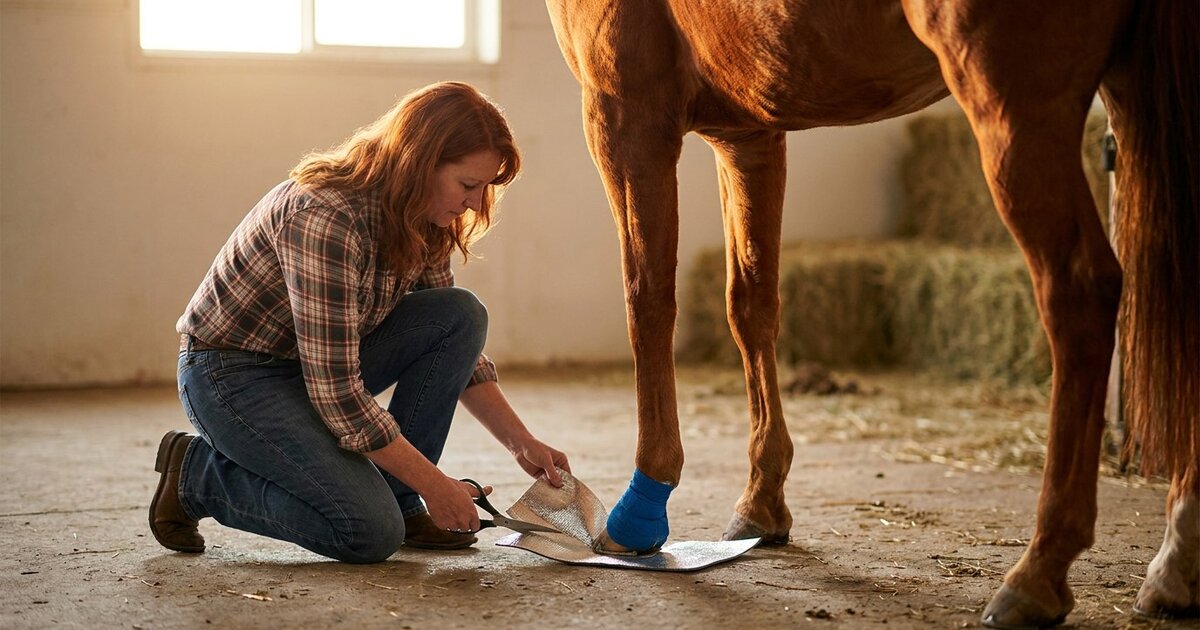
Animalintex pads: These pre-made poultice pads are the gold standard for convenience. Cut to fit the sole, wet with warm water, apply the white (medicated) side against the sole, and bandage in place. They contain boric acid and tragacanth, which draw infection toward the surface. Replace every 12-24 hours.
Ichthammol (drawing salve): A thick, black, tar-like ointment that draws moisture and encourages drainage. Apply a thick layer to the sole, cover with gauze or a disposable diaper (yes, newborn diapers work perfectly for this), and bandage. Ichthammol is cheap, effective, and available at most farm supply stores for $6-10 a tin.
Homemade poultice: A paste of Epsom salts and betadine scrub (not solution) packed into the sole and covered with a diaper and duct tape boot works in a pinch. Not as elegant as Animalintex, but functional.
Step 4: Bandage and Protect
The poultice needs to stay in place and the sole needs to stay clean. Options for securing everything:
Duct tape boot: Layer duct tape across the bottom of the hoof over the poultice, extending it up the hoof wall slightly. Cheap but wears through quickly in turnout.
Rubber soaking/treatment boot: EasySoaker, Davis boot, or similar. Most convenient for repeated treatments. Costs $20-40 but reusable.
Vetrap and duct tape combo: Wrap vetrap around the hoof over the poultice, then cover the bottom with duct tape. Secure above the coronary band but not too tight around the pastern.
Change the poultice and bandage every 12-24 hours. Inspect the sole each time for signs of drainage. When the abscess pops, you'll see dark, sometimes foul-smelling discharge. That's actually a great sign. Once it drains, the horse's lameness improves dramatically, often within hours.
Step 5: After Drainage
Once the abscess has drained, the goal shifts from drawing to protecting. Clean the drainage tract gently with dilute betadine solution. Pack the hole with cotton soaked in betadine or apply a thin layer of ichthammol. Continue bandaging for 2-3 days after drainage to prevent dirt and debris from entering the open tract.
The hole will grow out naturally over the next several weeks to months as the hoof grows. If the abscess blew out at the coronary band (a "gravel"), you'll see a disruption in the hoof wall as it grows down. This is cosmetic and typically resolves as the hoof replaces itself over 6-9 months.
Pain Management
Hoof abscesses hurt. A lot. While you're waiting for drainage, the horse may need pain relief. Phenylbutazone (bute) at 1-2 grams orally twice daily (consult your vet for exact dosing) is the most common choice. Some vets prefer flunixin meglumine (Banamine) for its visceral pain properties.
Here's a debate in the equine world: some practitioners advise against giving bute for abscesses because pain relief can mask the horse's ability to protect the foot, and the anti-inflammatory effect might slow the abscess from "coming to a head." There's logic there, but there's also logic in not letting a horse suffer unnecessarily. Discuss with your vet. My personal take: moderate pain relief is humane and appropriate while you're actively treating the abscess. Just don't use bute as a substitute for proper treatment.
When to Call the Vet
Most simple abscesses don't require veterinary intervention. But some do. Call the vet if:
The lameness doesn't improve within 7-10 days despite consistent soaking and poulticing.
The horse develops a fever (over 101.5°F rectal temperature in an adult horse at rest).
Multiple feet are involved. This could indicate laminitis rather than simple abscesses.
The abscess resulted from a nail puncture, especially in the central frog area.
Drainage is excessive or continuous beyond 2-3 days, which might indicate a deeper infection.
The horse has a history of chronic abscesses in the same foot, suggesting underlying hoof pathology, white line disease, or keratoma.
You're unsure of the diagnosis. When in doubt, get professional eyes on it.
Radiographs (X-rays) may be needed to rule out coffin bone infection (osteitis), foreign body, or other structural problems. For nail punctures, advanced imaging and even surgical exploration of the puncture tract may be necessary.
Preventing Hoof Abscesses
You can't prevent every abscess. But you can reduce the frequency:
Manage mud and moisture. Provide dry footing in at least part of the turnout area. Gravel pads around water troughs and gates help enormously.
Maintain regular farrier care. Every 6-8 weeks, no exceptions. A balanced hoof with strong walls and a healthy white line is the best defense.
Support hoof quality nutritionally. Biotin at 20mg per day has been shown to improve hoof wall growth and quality over 6-9 months. Zinc, copper, and methionine also support keratin production.
Keep paddocks clean. Pick up debris, nails from fence repairs, and wire. One nail in a paddock is all it takes.
Consider hoof boots or pads for horses with thin soles that work on rocky terrain.
Frequently Asked Questions
How long does a hoof abscess take to heal?
Once the abscess drains, most horses are dramatically better within 24-48 hours. Full resolution, where the drainage stops and the tract seals over, takes about 5-10 days. The hoof wall disruption from a coronary band blowout takes 6-9 months to grow out completely. But as far as soundness goes, most horses are back to work within 1-2 weeks of drainage.
Can I turn my horse out with an abscess?
In a small, clean, dry paddock, yes. You need to protect the bandaged foot from mud and debris. Deep mud, rocky ground, or large wet pastures will make treatment harder and contamination more likely. If you can't keep the hoof relatively clean in turnout, stall rest during the acute phase (3-5 days) is better.
Can a hoof abscess cause permanent damage?
Simple abscesses rarely cause lasting problems. However, a deep abscess that involves the coffin bone can cause osteitis (bone infection) requiring prolonged antibiotic treatment. Nail punctures into the navicular bursa or coffin joint can be career-ending or life-threatening without prompt, aggressive veterinary treatment. This is why puncture wound location matters so much.
My horse keeps getting abscesses in the same foot. Why?
Recurring abscesses in one foot suggest an underlying issue. Possibilities include chronic white line disease, a keratoma (a benign growth inside the hoof wall that creates a pocket for bacteria), poor hoof conformation, or thin soles. Radiographs and a thorough hoof exam by your vet and farrier working together can usually identify the root cause. Don't just keep treating repeat abscesses without investigating why they keep happening.
Is it true that abscesses are more common in spring?
Yes. Spring's freeze-thaw cycles and mud-to-dry transitions cause the hoof to expand and contract repeatedly, opening micro-pathways for bacteria. Fall is another peak season for the same reason. Horses that live in consistently dry or consistently wet conditions actually get fewer abscesses than those dealing with constant wet-dry fluctuations.
📝 Explore more in our Courses. Check it out here.
Sources
- American Association of Equine Practitioners. "Hoof Abscesses." aaep.org
- Merck Veterinary Manual. "Subsolar Abscess in Horses." merckvetmanual.com
- Colorado State University Veterinary Teaching Hospital. "Hoof Abscess Management." vetmedbiosci.colostate.edu
- O'Grady, Stephen E. "A Fresh Look at the Hoof Abscess." Equine Veterinary Education, 2019.
Last reviewed: April 2026