Horse Leg Injuries and Rehab: A Complete Owner's Guide |
You run your hand down the front leg on a Tuesday morning and something is different. A little fill. A little warmth. Maybe your horse is fine at the walk, so you tell yourself it's a stocked-up leg from standing in overnight. Then you trot him out and your stomach drops.
Almost every horse owner has that morning eventually. What happens next, over the following nine to twelve months, depends far more on how you handle the rehab than on any injection, boot, or supplement you buy in the first week.
Quick Answer
The most common horse leg injuries are soft tissue injuries to the flexor tendons and suspensory ligament, followed by wounds, splints, and joint problems. Tendon and ligament injuries typically require 9 to 12 months of controlled, veterinarian-guided rehabilitation to heal, because these structures have poor blood supply and heal with scar tissue rather than regenerating. Stall rest alone is not rehabilitation. Progressive, controlled exercise, adjusted by serial ultrasound, is what determines whether a horse returns to work or re-injures.
What Is Actually in a Horse's Lower Leg
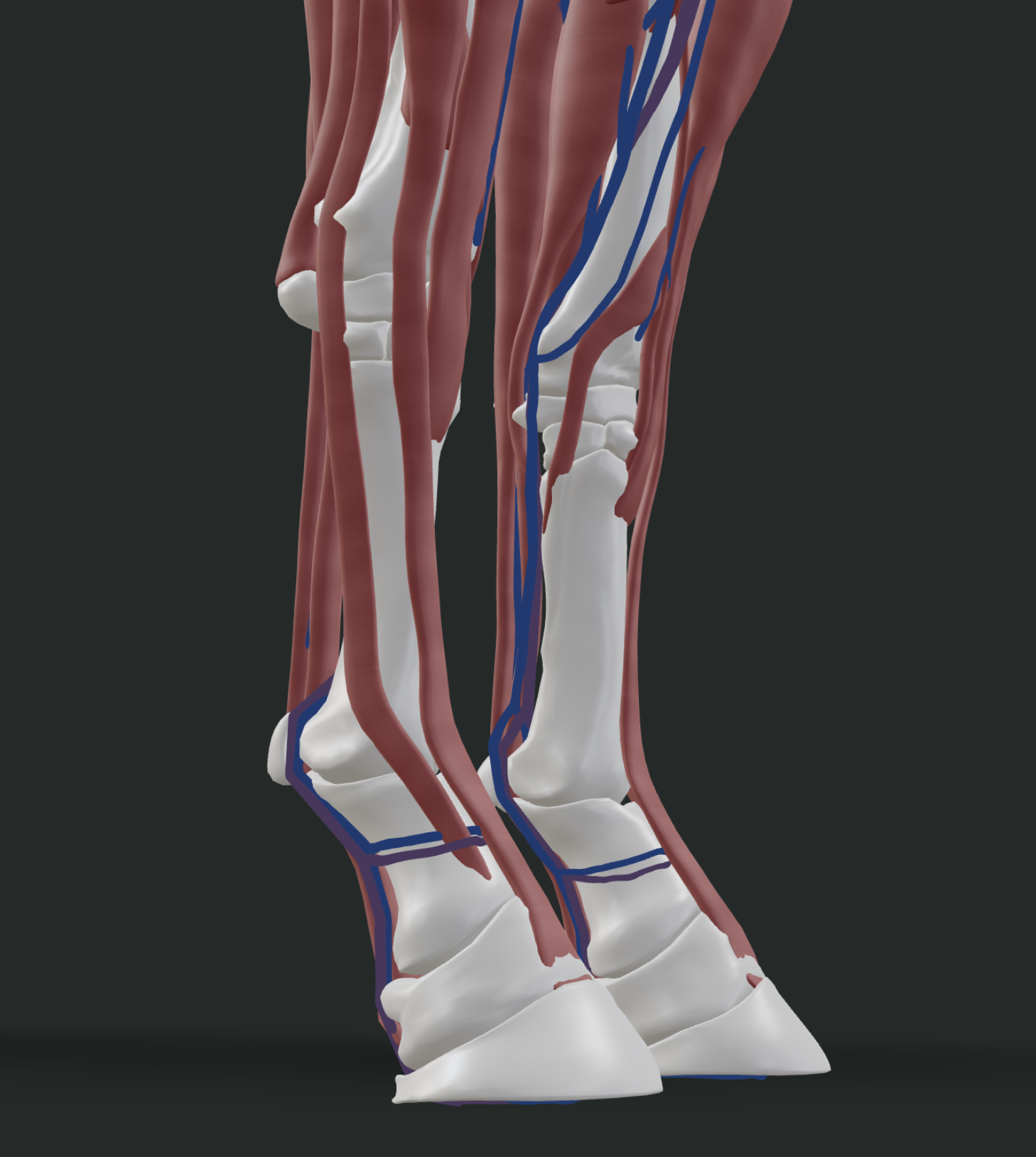
Before you can understand why these injuries happen, you have to understand what the horse is standing on. Below the knee and the hock, a horse has no muscle at all. What you feel under the skin is bone, tendon, ligament, joint capsule, and a thin layer of skin over the top of it.
That is a remarkable piece of engineering. It makes the lower limb light, which lets the horse swing it fast, which is how a prey animal outruns things. But it also means there is almost no cushioning. Muscle absorbs shock. Tendon does not. Tendon stores and returns energy like a spring, and springs eventually fatigue.
The structures that carry the load are:
- The superficial digital flexor tendon (SDFT), running down the back of the cannon bone. It supports the fetlock and stores elastic energy at speed.
- The deep digital flexor tendon (DDFT), running beneath the SDFT, continuing down and around the navicular bone to attach to the coffin bone.
- The suspensory ligament, running down the back of the cannon bone beneath the flexor tendons, then splitting into two branches that attach to the sesamoid bones at the back of the fetlock. It is the structure that stops the fetlock from dropping to the ground under load.
- The check ligaments, which limit how far the flexor tendons can stretch.
- The cannon bone and splint bones, the weight-bearing column.
Add one more fact and the picture is complete: the forelimbs carry roughly 60 percent of a horse's body weight. That is why the front legs break down more often than the hinds.
You can see all of these structures layered on top of each other in the Inside The Equine 3D explorer, where you can strip the muscle layer back and watch the flexor tendons and suspensory ligament run down the back of the cannon bone in three dimensions. It is the fastest way to understand why a "leg injury" is never just a leg injury. It is a specific structure, and which one it is changes everything about the prognosis.
The Most Common Horse Leg Injuries
Bowed tendon (SDFT tendinitis)
The classic. Overstretching tears collagen fibers inside the SDFT, blood and fluid fill the space, and the back of the cannon bone takes on the convex "bow" shape that gives the injury its name. The leg is hot, swollen, firm, and painful to press. In racehorses, the overwhelming majority of tendon and ligament injuries involve the SDFT.
A bow is the extreme end of a spectrum. Most horses do not go straight to a full bow. They pass through minor strains and low-grade tendinitis first, which is exactly the stage most owners miss.
Suspensory ligament injury (desmitis)
Suspensory injuries are the sport horse's version of the racehorse's bowed tendon. They can occur at the origin high up under the knee or hock (proximal suspensory desmitis), in the body, or in the branches down near the sesamoids.
The cruel part is that suspensory injuries frequently show no visible swelling, particularly proximal ones, because the ligament sits deep. A horse can have a significant suspensory lesion and a leg that looks entirely normal. Research on Thoroughbred racehorses puts suspensory ligament injury prevalence somewhere in the range of 3.6 to 10 percent, though that figure varies by population and study.
Deep digital flexor tendon injury
Less common than SDFT injury and often harder to find, because a good portion of the DDFT sits inside the hoof capsule where ultrasound cannot reach it. These frequently require MRI to diagnose properly and are a common finding in horses with chronic heel pain.
Splints
Inflammation of the interosseous ligament between the splint bone and the cannon bone, usually from concussion, a blow, or hard work in young horses. It produces heat, pain, and eventually a firm bony lump. Splints are usually far less serious than they look. The lump is often permanent and cosmetic once it has cooled and settled, but a hot, painful splint is an active injury and needs rest. See the conditions entry on splints for the full clinical picture.
Lower limb wounds
The lower leg is a bad place to get cut. There is little soft tissue, blood supply is poorer than higher up the body, and every joint movement pulls at the healing edges. Add bedding and mud and you have a contamination problem. This is why lower limb wounds are the ones that turn into proud flesh, and why a wound anywhere near a joint or tendon sheath is an emergency, not a "watch it for a few days" situation.
Cellulitis
A bacterial infection of the tissue under the skin, usually entering through a scratch or a case of scratches that nobody thought twice about. The leg blows up dramatically, often from the pastern upward, and it is hot and extremely painful. Cellulitis is one of the few leg problems where the swelling really is the emergency. It moves fast and it needs a vet the same day.
Fractures
Merck's guidance on suspected fractures is worth committing to memory. Suspect one if you hear a loud crack, if the horse is suddenly non-weight-bearing, if the limb hangs at an abnormal angle, or if it is visibly unstable. Do not move the horse. Call your vet.
Tendon Versus Ligament: The Difference That Matters
Owners use these words interchangeably. Vets do not, and the distinction changes the rehab plan.
| Tendon | Ligament | |
|---|---|---|
| Connects | Muscle to bone | Bone to bone |
| Function | Transmits muscular force, stores elastic energy | Stabilizes joints, limits movement |
| Key examples in the leg | SDFT, DDFT | Suspensory ligament, check ligaments, collateral ligaments |
| Injury term | Tendinitis / tendinopathy | Desmitis |
| Visible swelling | Usually yes, often dramatic | Often none, especially proximal suspensory |
| Primary imaging | Ultrasound | Ultrasound; MRI for proximal and foot lesions |
How Leg Injuries Get Diagnosed
A proper workup usually runs in this order: a lameness exam with the horse trotted in hand and often on a circle, palpation of the limb for heat, pain, and swelling, flexion tests, and diagnostic nerve blocks to localize the pain to a region.
Only then does imaging happen, and it happens where the blocks pointed. Ultrasound is the primary tool for soft tissue and gives a baseline measurement of lesion size, cross-sectional area, and fiber pattern. Radiographs rule out bony involvement. MRI comes in when the lesion is inside the hoof or the picture does not add up.
That baseline ultrasound matters enormously, and it is worth understanding why. Every later decision in the rehab is made by comparing new images against it. Skip the baseline and you spend the next nine months guessing.
What Rehabilitation Actually Means
Here is the thing most owners get wrong, and it is the single most expensive mistake in this entire article.
Stall rest is not rehabilitation. A tendon left completely unloaded heals into disorganized scar tissue with poor fiber alignment and poor strength. It is the loading that tells the new collagen which way to line up.
Healing runs through three phases. First inflammation, in roughly the first one to two weeks, where the priority is limiting further damage. Then proliferation, where cells lay down type III collagen: fast to produce, weaker and less organized than the original. Then remodeling, a long slow phase where that type III collagen is gradually converted toward stronger, better-aligned type I collagen. That remodeling phase is where controlled exercise does its work, and it is measured in months, not weeks.
A typical veterinary rehabilitation program follows this shape:
| Phase | Roughly when | What it looks like |
|---|---|---|
| Acute | Weeks 1 to 2 | Confinement, cold therapy, support bandaging, baseline ultrasound |
| Early controlled exercise | Weeks 2 to 12 | Stall confinement with short daily hand-walking, increasing gradually |
| Trot introduction | Around months 3 to 5 | Straight lines only, firm level footing, guided by recheck ultrasound |
| Canter introduction | Around months 6 to 9 | Only after imaging supports it. The highest re-injury window |
| Return to work | 9 to 12 months | Final ultrasound before full athletic loading |
Two enormous caveats on that table. These timelines are illustrative, not a prescription. Your vet builds the actual program from your horse's specific lesion, and the whole point of serial ultrasound (commonly repeated at roughly 60-day intervals) is that the program gets adjusted to what the tissue is actually doing rather than to a calendar.
And the numbers are why people fail. Most horses look and feel sound long before the tendon is strong. A horse can be jogging happily and pain-free at four months with a lesion that would tear again the first time you asked for a canter. The horse's comfort is not the metric. The ultrasound is.
Why Re-Injury Is the Real Enemy
Repaired tendon is not new tendon. It is scar, and scar is biomechanically inferior to the original tissue. That is the fundamental, unglamorous truth of soft tissue healing in horses, and it is why re-injury rates for flexor tendon injuries are high enough that clinicians treat the return-to-canter phase as the most dangerous moment in the entire process.
The two most common reasons a rehab fails are worth stating plainly. The first is non-compliance: the owner or the horse cutting the controlled exercise program short. The second is a missed secondary problem. A horse with sore hocks will not rehabilitate a front suspensory well, because he keeps unloading the hind end and overloading the front, no matter how perfectly you follow the walking schedule.
If you want a broader map of how lameness gets localized in the first place, the equine lameness guide covers the diagnostic process in more depth, and the horse leg anatomy guide walks through the structures themselves. For upper limb problems that present as vague hind-end lameness, start with hock injuries.
And if your horse is in a rehab program right now, it is worth spending ten minutes with the 3D explorer's skeletal and muscular layers looking at the exact structure your vet named. Owners who can picture the lesion tend to follow the controlled exercise program better, because the timeline stops feeling arbitrary and starts making sense.
Frequently Asked Questions
How long does a horse leg injury take to heal?
Most tendon and ligament injuries take 9 to 12 months to heal completely. Mild to moderate cases may allow ridden walking around 3 to 4 months, but full return to athletic work is generally not attempted before 9 months. Wounds, splints, and bruises heal much faster, but soft tissue injuries are measured in seasons, not weeks.
Can a bowed tendon fully heal?
A bowed tendon heals, but it heals with scar tissue rather than regenerating original tendon. Many horses return to full work. The repaired tendon is permanently thicker and biomechanically weaker than the original, which is why re-injury risk stays elevated and why the controlled exercise program cannot be rushed.
Should I cold hose a swollen leg?
Cold therapy is a standard first response to acute soft tissue injury and is commonly recommended alongside support bandaging in the first days. It is not a substitute for a diagnosis. If a leg is hot, swollen, and the horse is lame, cold hosing while you wait for your vet is reasonable. Cold hosing instead of calling your vet is not.
Why does my horse's suspensory injury have no swelling?
Proximal suspensory ligament injuries sit deep in the limb, high up under the knee or hock, and frequently produce no visible swelling at all. This is why suspensory injuries are so often caught late. Lameness without swelling is not reassuring. It is a reason to have the leg blocked and scanned.
Is stall rest enough to heal a tendon injury?
No. Complete rest produces poorly organized scar tissue with weak fiber alignment. Progressive, controlled loading is what drives the collagen to remodel into a stronger, better-aligned structure. This is why veterinary rehabilitation protocols are built around graduated exercise rather than pure confinement.
What is the difference between a splint and a bowed tendon?
A splint is inflammation and eventual bony growth involving the splint bone and the ligament attaching it to the cannon bone. It sits on the side of the leg and the lump is hard. A bowed tendon involves the flexor tendons at the back of the cannon bone, the swelling is soft and diffuse in the acute phase, and the prognosis is far more serious.
Want a complete guide to leg injury rehab?
Our Lameness course covers injury types, recovery timelines, and rehabilitation protocols.
Explore the Lameness CoursePro members only. Try Pro free for 7 days.