Horse Neck and Cervical Spine Anatomy Explained
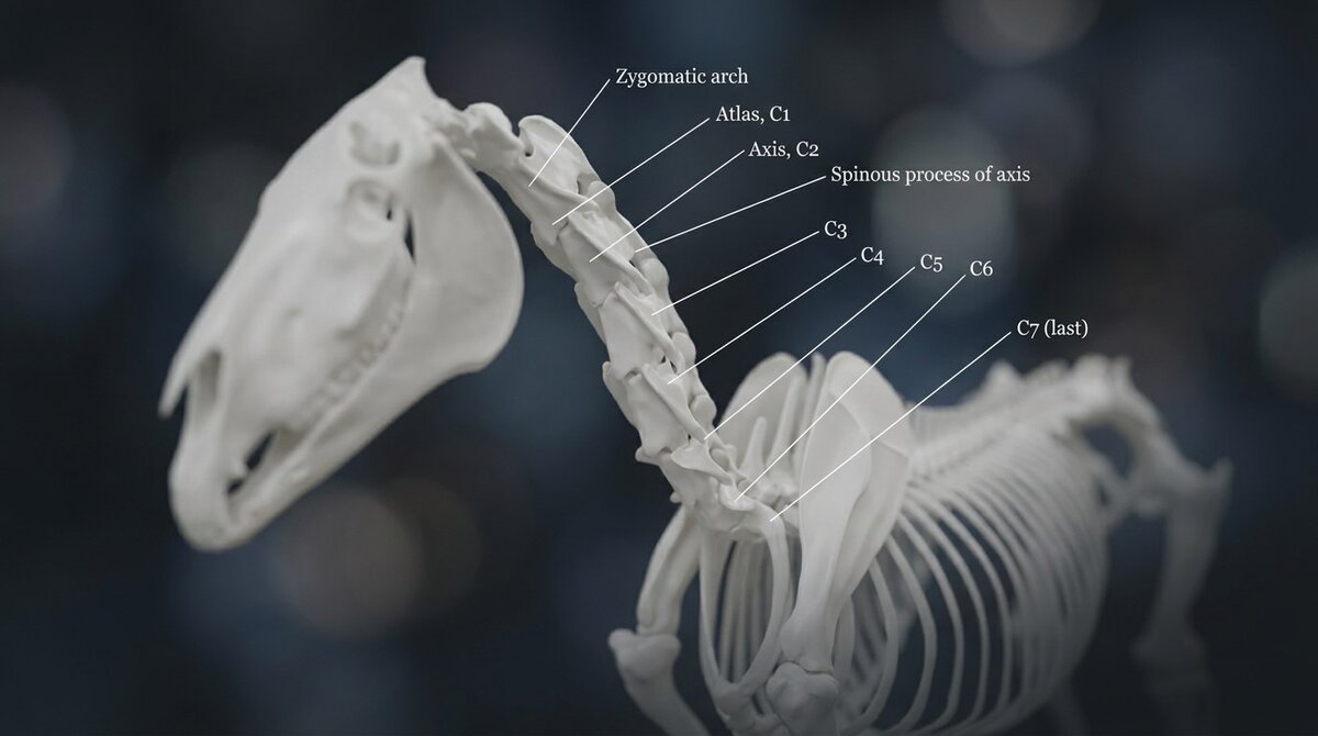
Horse Neck and Cervical Spine Anatomy
Ask most people to point to a horse's neck bones and they will draw a line along the top of the crest, following the mane. They will be wrong by several inches, and that misunderstanding is at the root of a great deal of confusion about how horses carry themselves.
The horse's neck does not run where it looks like it runs. Knowing where it actually is changes how you think about everything from bending to bitting to why a horse braces.
Seven Vertebrae, Same as a Giraffe
Nearly every mammal has exactly seven cervical vertebrae. A mouse has seven. A human has seven. A giraffe has seven. The difference is the size of each bone, not the number of them. It is one of the most conserved patterns in mammalian anatomy, and the horse follows it.
Those seven bones are labelled C1 through C7, from the skull down toward the chest. But they are not all the same, and the differences matter.
The Atlas and the Axis
The first two vertebrae are specialists.
C1, the atlas, is the ring of bone that connects directly to the skull. It is named for the figure who held up the world, because it holds up the head. The joint between the skull and the atlas is the "yes" joint. It is what lets the horse nod, raising and lowering the muzzle.
C2, the axis, sits just below and is the longest of the cervical vertebrae. The joint between the atlas and the axis is the "no" joint. It is what lets the horse rotate the head, the pivoting motion you see when a horse tips its nose to the side.
Together, these top two joints give the head most of its fine mobility. The research on how the equine neck moves shows that these first two joints move considerably more than the joints further down. The top of the neck is where the delicate, precise movement lives.
Where the Neck Actually Is
Now the part that reorganizes everything.
The cervical spine does not run along the crest. It runs in an S-shape. From the poll it angles down and back, dips to its lowest point deep in the base of the neck just in front of the chest, and then the first thoracic vertebrae rise again into the withers. The lowest part of the working neck is well below the crest, sitting closer to the level of the point of the shoulder than most people expect.
Why does this matter? Because when a rider talks about a horse "lifting the base of the neck" or "telescoping the neck forward," they are talking about this hidden lower curve, not the visible topline. A horse can have a beautifully arched crest and a dropped, braced base of the neck at the same time. The crest is not the spine.
Seeing this is genuinely hard from a diagram and easy in three dimensions. Turn on the skeletal layer in the 3D explorer and look at the neck from the side, and the S-curve becomes obvious. Then the whole conversation about neck posture starts to make sense.
The Facet Joints
Between each pair of vertebrae, on each side, sits a small joint called an articular process joint, or facet joint. These are gliding joints, and their job is to guide and permit the bending and turning of the neck.
They matter clinically because, like any joint, they can develop arthritis. Facet joint arthritis in the neck can cause stiffness, resistance to bending one way, pain, and in some cases pressure on the nerve roots that exit between the vertebrae. Because those nerve roots feed the neck and the forelimbs, neck problems can sometimes show up as vague forelimb lameness or an unwillingness to work in a particular direction. This is one of the reasons a horse that "won't bend right" or "feels off in front" sometimes turns out to have a neck issue rather than a leg issue.
The Nuchal Ligament
The horse does not hold its heavy head up with muscle alone. That would be exhausting. Instead it uses a large elastic structure called the nuchal ligament, which runs from the back of the skull along the top of the neck and connects into the ligament system that continues down the back to the sacrum.
Think of it as a suspension cable. When the horse lowers its head to graze, the nuchal ligament stretches and stores energy. When the horse raises its head, that stored energy helps lift it back up. It is a passive, elastic support system that lets the horse carry a heavy head on a long neck without burning muscle the whole time.
This is also why encouraging a horse to stretch long and low is genuinely valuable work. It loads and lengthens this whole system along the topline, from poll to tail.
The C6 and C7 Question
In recent years a lot of anxious conversation has surrounded the shape of the sixth and seventh cervical vertebrae, under names like ECVM and ECCMV (equine complex vertebral malformation, equine caudal cervical morphologic variation). The short version is that some horses have small variations in the bony processes at the base of the neck.
Here is what the current research actually says, and it is reassuring. A 2026 study of 200 clinically sound warmblood sport horses found these variations present in roughly a third of them, found no association between the variation and competition results, and concluded that it appears to be a common anatomical characteristic rather than a disease. The variation was there in horses competing perfectly well.
That does not mean the base of the neck never causes problems. It means that finding one of these variations on a radiograph is not, on its own, a diagnosis or a reason to panic. It is a common finding whose significance has to be judged case by case, by a vet, in the context of the actual horse. It is worth knowing the term exists so that if it comes up you do not assume the worst.
Why It All Connects
The neck is the horse's balancing pole and its steering. It carries a heavy head at the end of a long lever, it houses the spinal cord feeding the front half of the body, and it is the first thing that changes when a horse braces or relaxes. Understanding that the working spine sits low, that the top joints do the fine movement, and that the whole thing is slung on an elastic ligament makes almost everything about neck posture and training more intelligible.
The horse anatomy reference puts the neck in the context of the whole spine, and the 3D explorer is the fastest way to see the S-curve for yourself.
How many bones are in a horse's neck?
A horse has seven cervical vertebrae, labelled C1 through C7. This is the same number found in almost all mammals, including the giraffe. The bones differ in size and shape along the neck, but the count is seven.
Where is a horse's neck bone actually located?
The cervical spine runs in an S-shaped curve, not along the top crest where the mane sits. It dips to its lowest point deep at the base of the neck near the chest before rising into the withers. The working spine sits well below the visible crest, which is why the crest is a poor guide to what the neck is doing.
What are the atlas and axis in a horse?
The atlas (C1) is the first vertebra, connecting to the skull and allowing the head to nod up and down. The axis (C2) is the second and longest vertebra, allowing the head to rotate side to side. Together they provide most of the head's fine mobility.
Is C6/C7 malformation in horses serious?
Not necessarily. Variation in the shape of the sixth and seventh cervical vertebrae is common. A 2026 study of clinically sound warmblood sport horses found it in roughly a third of them with no effect on competition results, concluding it is often a normal anatomical variant rather than a disease. Its significance in any individual horse should be judged by a vet.
Can neck problems cause front leg lameness in horses?
They can. The nerve roots that supply the forelimbs exit between the cervical vertebrae, so arthritis or other changes in the neck's facet joints can sometimes produce forelimb lameness or an unwillingness to bend or work in one direction. This is why a persistent forelimb issue is sometimes traced back to the neck.
Frequently Asked Questions
How many bones are in a horse’s neck?
Seven cervical vertebrae (C1–C7), the same number found in nearly all mammals including giraffes. Size, not number, accounts for length differences.
Where is a horse’s neck actually located?
The cervical spine forms an S-curve that dips low at the base of the neck near the chest, not along the crest. The working spine sits well below the visible topline.
What are the atlas and axis?
C1 (atlas) allows nodding (“yes”); C2 (axis) allows rotation (“no”). These two joints provide most of the head’s fine mobility.
Is C6/C7 variation in horses a serious problem?
Usually not. A 2026 study of sound warmbloods found the variation in roughly one-third of horses with no impact on performance; significance must be judged case-by-case by a veterinarian.
Quick Reference Table
| Vertebra | Common Name | Key Features | Clinical Significance |
|---|---|---|---|
| C1 | Atlas | Wing-shaped, no body; articulates with skull | Allows yes (nodding) motion; fractures are usually fatal |
| C2 | Axis | Large dens (peg) projects into C1 | Allows no (rotation) motion; trauma can cause wobbler signs |
| C3 | (none) | Typical cervical shape, short spinous process | Articular facet arthritis common in older horses |
| C4 | (none) | Similar to C3 | Mid-neck stiffness often involves C3-C5 |
| C5 | (none) | Transitional features begin | Narrowing of vertebral canal can compress spinal cord |
| C6 | (none) | Prominent ventral lamina (crest on underside) | Landmark for ultrasound; common site for cervical stenosis |
| C7 | (none) | Short, transitions to thoracic spine | Articulates with first rib; injury affects forelimb gait |