Horse Skull and Head Anatomy: What's Behind the Face
Horse Skull and Head Anatomy: What's Behind the Face
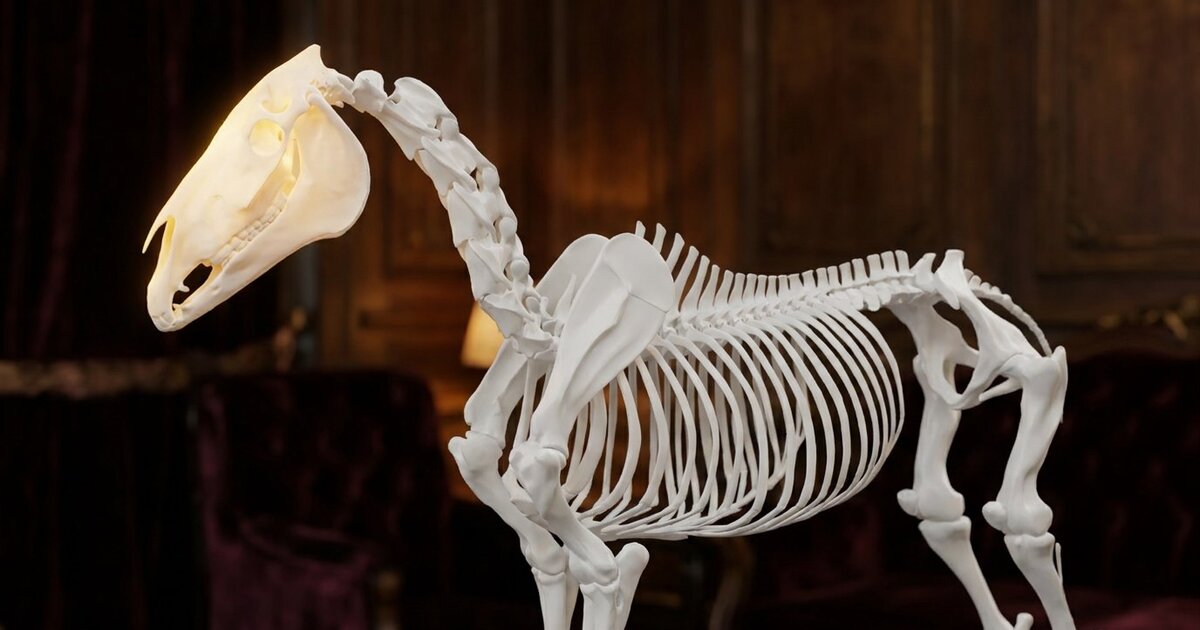
Pick up a horse's skull and the first thing that surprises you is how light it is. For a head that long, it weighs almost nothing, and the reason is that most of it is not bone at all. It is air.
That single fact, that the horse's head is mostly hollow, explains why a tooth problem can turn into a face full of infection, why a horse can crack a bone in its head and mostly shrug, and why the whole structure works the way it does.
The Skull Is Built From Many Fused Bones
The horse skull is not one bone. It is a set of them, knitted together at seams called sutures. In a young horse these sutures are visible and slightly mobile. As the horse matures they fuse, and in an adult most of the skull is effectively a single rigid structure.
The significant exception is the mandible, the lower jaw. It is the one large part of the head that moves, hinging at the temporomandibular joint just below the ear and swinging in the wide side-to-side grinding motion that a grazing animal needs. Watch a horse eat hay and you are watching the mandible do the one job the rest of the skull cannot.
You can see how the bones fit together, and how the jaw articulates, in the Inside The Equine 3D explorer with the skeletal layer on the head.
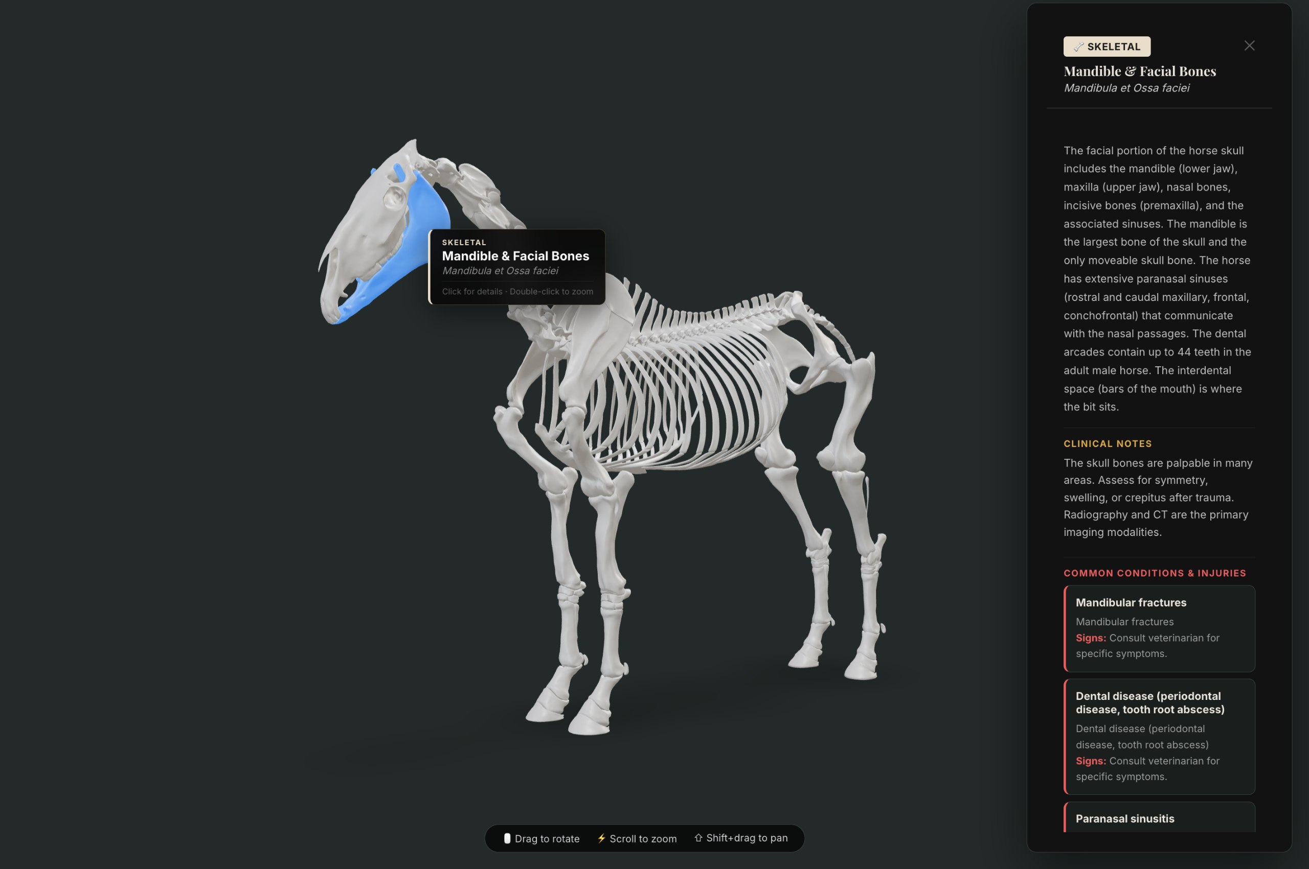
The mandible and facial bones in the Inside The Equine 3D Explorer. Tap any structure to see clinical notes and linked conditions.
The Head Is Mostly Air
Here is the part that reframes everything. Running through the bones of the horse's face is a system of air-filled chambers called the paranasal sinuses. They are large, they are interconnected, and they take up a great deal of the space you would assume was solid.
The sinuses do several useful things. They lighten the head, which matters enormously for an animal that spends its life balancing a long heavy skull on the end of a long neck and swinging it around. They warm and humidify the air the horse breathes. And they give the voice its resonance.
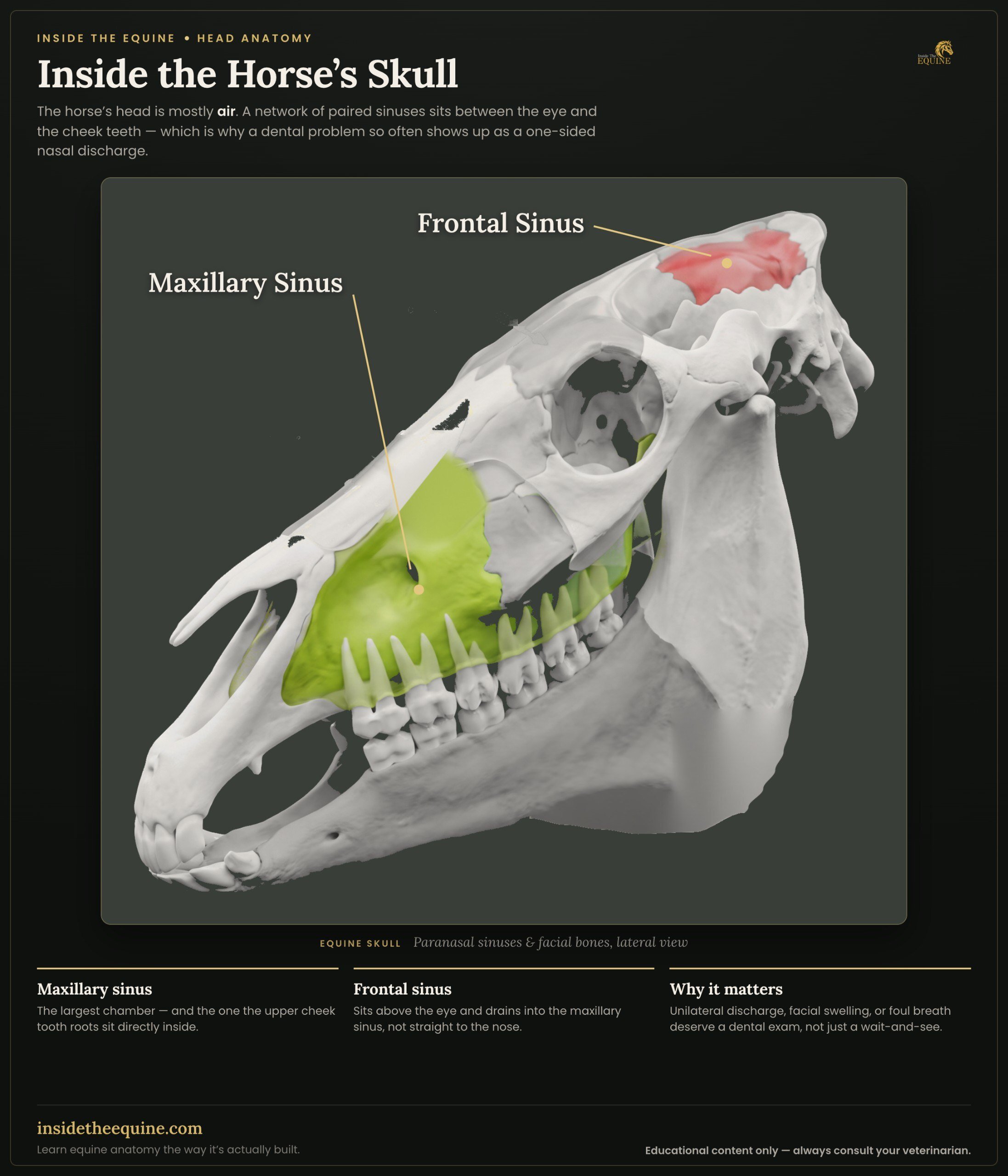
The largest and most clinically important are the maxillary sinuses, the big chambers in the cheek region on each side of the face. Remember those. They are where the trouble happens.
The paranasal sinuses and facial bones of the horse skull, lateral view. Note how the upper cheek tooth roots sit directly inside the maxillary sinus. From Inside The Equine.
Why Dental Problems Become Sinus Problems
This is the single most useful thing to understand about the horse's head, and it follows directly from the anatomy.
A horse's cheek teeth are enormous. The crown you can see in the mouth is only a fraction of the whole tooth. The rest, the reserve crown and the roots, extends far up into the bone of the upper jaw. And in the upper jaw, the back cheek teeth project their roots directly into the floor of the maxillary sinus.
Sit with that for a second. The roots of the upper back teeth are sitting inside the air-filled sinus cavity, separated from it by only a thin plate of bone, or sometimes by nothing at all.
The consequence is unavoidable. When one of those upper cheek teeth develops an infection at the root, the infection has a very short distance to travel before it is inside the sinus. A tooth root abscess becomes a sinus infection, and the horse presents with a one-sided nasal discharge, often smelly, that no amount of treating "a cold" will fix, because it was never a cold. It was a tooth.
This is why a persistent, one-sided, foul-smelling nasal discharge in a horse is a reason to have the teeth examined, not just the nose. The nose is the symptom. The tooth is often the cause. Understanding the horse's teeth and dental anatomy makes the whole picture click into place.
The Guttural Pouches
Now to one of the strangest structures in the entire animal, and one that horses share only with a few close relatives.
The guttural pouches are two large air-filled sacs, one on each side, tucked at the back of the head between the base of the skull and the throat. They are outpouchings of the eustachian tubes, the tubes that connect the middle ear to the throat, and in the horse those tubes balloon out into these substantial cavities.
Nobody is entirely certain what they are for. The leading idea is that they help cool the blood traveling up to the brain, because of what passes through them, which brings us to why they matter medically.
Running along the walls of the guttural pouches are major arteries and important nerves, including vessels carrying blood to the brain and nerves controlling swallowing and the larynx. This makes the guttural pouches a genuinely dangerous piece of real estate. A fungal infection that takes hold in a guttural pouch can erode one of those arteries and cause sudden, severe, life-threatening bleeding from the nose. Infections here can also damage the nerves and interfere with swallowing.
You do not need to be able to diagnose any of this. You need to know the guttural pouches exist, that they sit at the back of the head, and that they are one more reason a nosebleed or a swallowing problem in a horse is worth a vet's attention rather than a wait-and-see.
The Eyes and the Long Face
The horse's eyes sit high and to the sides of that long head, and the length of the face is part of what makes their vision work. With eyes set where they are, a grazing horse can keep its muzzle down in the grass and still see almost all the way around itself, watching for anything that might be watching it.
The eye sockets, the orbits, are complete rings of bone in the horse, which gives the large eyeball solid protection. The horse's eye is the largest of any land mammal, and the anatomy of the head is built around keeping those two big eyes up, wide, and scanning. The equine eye is worth its own look, but the point here is that the shape of the skull and the placement of the eyes are the same story: a prey animal built to see a threat coming.
Why the Structure Makes Sense
Step back and the whole head reads as one design. Long, so the eyes sit high while the mouth grazes. Light, because it is full of air rather than bone, so that long lever is not a burden to carry and swing. Deep-rooted grinding teeth, because a life of chewing abrasive grass wears teeth down and the tooth has to keep feeding more crown up into the mouth. And a jaw built to move side to side, because that is how you grind, rather than up and down, which is how a predator bites.
The muscular system of the horse overlaid on the skeleton, showing how the head and neck muscles connect to the trunk. From the Inside The Equine 3D Explorer.
Every strange feature, the air-filled face, the teeth buried in the sinus, the pouches at the back of the skull, is a solution to the problem of being a large animal that survives by eating grass and not being eaten. Seeing it laid out in three dimensions is the fastest way to make it stick, and the 3D explorer lets you turn the head, strip it back, and look.
Why does my horse have a one-sided nasal discharge?
A one-sided nasal discharge, particularly if it is foul-smelling, is frequently caused by a problem with an upper cheek tooth rather than a problem with the nose. The roots of the upper back teeth sit inside the maxillary sinus, so a tooth root infection can drain into the sinus and out through that nostril. It should be examined by a vet, and the examination needs to include the teeth.
What are guttural pouches in horses?
The guttural pouches are two large air-filled sacs at the back of a horse's head, formed as outpouchings of the eustachian tubes. They are found only in horses and a few related species. Major arteries and nerves pass through them, which is why infections of the guttural pouches can be serious, occasionally causing severe nosebleeds or swallowing problems.
Why is a horse's head so long?
The length of the head allows the eyes to sit high and to the sides while the muzzle reaches the ground to graze, so the horse can watch for predators while eating. The long face also houses the large grinding cheek teeth and the extensive air-filled sinuses that keep the whole structure light.
Are a horse's teeth connected to its sinuses?
Yes, in the upper jaw. The roots of the upper cheek teeth project up into the maxillary sinuses, separated by only a thin plate of bone. This close relationship is why dental infections in the upper back teeth commonly cause sinus infections in horses.
How many bones are in a horse's skull?
The horse skull is made up of numerous bones joined at sutures, most of which fuse as the horse matures into what is effectively a single rigid structure. The mandible, or lower jaw, remains separate and mobile. Sources count the individual bones differently depending on how fused elements are treated.
Quick Reference Table
| Structure | Location | Function | Clinical Relevance |
|---|---|---|---|
| Frontal Bone | Forehead, between eyes | Protects brain, forms frontal sinus | Sinus trephination site for treating sinusitis |
| Maxilla | Upper jaw | Houses upper teeth roots, forms maxillary sinus | Tooth root infections drain into sinus causing one-sided nasal discharge |
| Mandible | Lower jaw (only mobile skull bone) | Houses lower teeth, allows chewing | Fractures from kicks; important for bit placement |
| Nasal Bones | Bridge of nose | Protect nasal passages | Fragile; easily fractured by trauma, causing epistaxis |
| Temporal Bone | Side of skull, behind eye | Contains TMJ, protects inner ear | TMJ problems can cause difficulty chewing |
| Hyoid Apparatus | Suspended below skull base | Supports tongue and larynx | Fracture causes difficulty swallowing; connected to guttural pouches |
| Guttural Pouches | Behind skull, below ear | Air-filled sacs (function debated; may cool blood) | Fungal infection can erode arteries causing fatal hemorrhage |
| Paranasal Sinuses | Throughout upper face | Lighten skull weight | Sinusitis from dental disease is common |
Frequently Asked Questions
How many bones are in a horse's skull?
The horse skull is made up of 34 individual bones, though most of them fuse as the horse matures into what effectively functions as one rigid structure. The mandible (lower jaw) remains the only freely moving bone. Exact counts vary between sources because some fused elements can be counted as one or two bones.
Why is a horse's head so long compared to other animals?
The length serves two purposes. First, it allows the eyes to sit high and wide on the sides of the head so the horse can watch for predators while grazing with its muzzle on the ground. Second, the long upper jaw houses a row of large, deep-rooted grinding teeth needed for a lifetime of chewing abrasive forage.
What are the sinuses in a horse's head and why do they matter?
The paranasal sinuses are air-filled cavities within the bones of the face. They significantly reduce the weight of the horse's long head. The maxillary sinus is clinically important because the roots of the upper cheek teeth sit inside it, meaning a tooth root infection can become a sinus infection, producing a foul-smelling one-sided nasal discharge.
What are guttural pouches and why are they dangerous?
Guttural pouches are two large air-filled sacs unique to horses and a few related species, formed as outpouchings of the eustachian tubes. Major arteries and cranial nerves run along their walls. A fungal infection (guttural pouch mycosis) can erode an artery wall and cause sudden, life-threatening hemorrhage from the nose.
Can a horse get a sinus infection from a bad tooth?
Yes, this is one of the most common causes of sinusitis in horses. The roots of the upper cheek teeth project directly into the maxillary sinus. When a tooth root becomes infected or abscessed, bacteria spread into the sinus. The classic sign is a persistent, foul-smelling discharge from one nostril only.
Why does my horse have a bump on its forehead?
Young horses often have noticeable bumps on their foreheads from the developing frontal sinuses expanding as they grow. In adult horses, a new or growing bump over the sinus area could indicate a sinus cyst, tumor, or bone infection and should be evaluated by a veterinarian.
How does the horse's skull protect its brain?
The brain sits at the back and top of the skull, well protected by thick bone. The frontal region that looks like the "forehead" is actually mostly sinus space, not brain. This means a blow to the front of the face is more likely to fracture sinus bones than to cause brain injury, though any head trauma warrants veterinary assessment.