Equine Nervous System: How Horses Sense and React
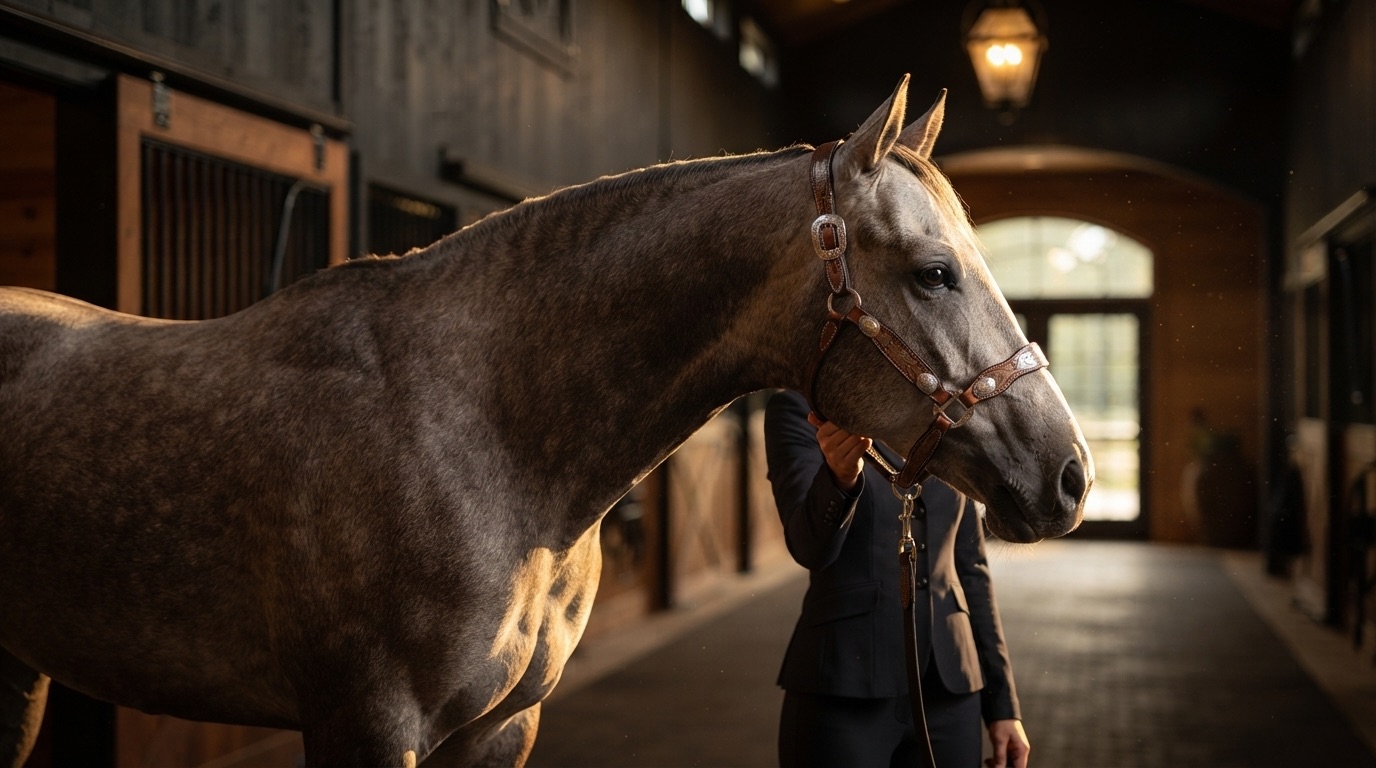
A horse spooks at a plastic bag forty feet away but stands perfectly still while you stitch up a wound on its leg. Makes no sense, right? Except it does, once you understand how the equine nervous system actually works. The wiring in a horse is built for survival first and everything else second. Their entire neurological architecture prioritizes speed of reaction over accuracy of interpretation, which is exactly why a 1,200-pound animal can lose its mind over a butterfly.
Quick Answer: The equine nervous system consists of the central nervous system (brain and spinal cord) and the peripheral nervous system (cranial nerves, spinal nerves, and the autonomic system). Horses have highly developed sensory pathways that prioritize rapid threat detection. Their large eyes provide nearly 350 degrees of vision, their ears rotate 180 degrees independently, and their skin contains one of the densest concentrations of tactile receptors of any large mammal. The autonomic nervous system governs the fight-or-flight response that defines so much of equine behavior.
The Central Nervous System: Command Center
The Equine Brain
A horse brain weighs roughly 600 to 700 grams. For perspective, that is about the size of a large grapefruit. Compared to body mass, it is relatively small, but raw size tells you almost nothing about function. What matters is how the brain is organized, and in horses, the allocation is telling.
The cerebrum handles higher processing, learning, and voluntary movement. In horses, the cerebral cortex is less convoluted than in primates but more developed than in many other ungulates. The areas devoted to sensory processing, particularly vision and hearing, are proportionally enormous. The olfactory bulbs are also substantial. Horses rely on smell more than most riders realize. A stallion can detect a mare in heat from remarkable distances, and horses routinely use scent to identify herd members, detect predators, and assess food quality.
The cerebellum sits at the back of the brain and coordinates movement and balance. Given that a horse must coordinate four limbs at various gaits while carrying a rider over uneven terrain, the cerebellum pulls heavy duty. Damage here shows up as ataxia, that drunken, swaying walk that sends every horse owner into a panic. Equine protozoal myeloencephalitis (EPM) and cervical vertebral malformation (wobblers syndrome) both target this region with devastating effect.
The brainstem connects the brain to the spinal cord and manages the functions you never think about until they go wrong: heart rate, respiration, swallowing, consciousness. It is the most primitive and most essential part of the brain. Damage to the brainstem is almost always catastrophic.
The Spinal Cord
Running from the brainstem through the vertebral canal to approximately the first sacral vertebra, the spinal cord is the information superhighway of the body. It carries motor commands down from the brain and sensory information up. In an average adult horse, the spinal cord is about 200 centimeters long and roughly 2 centimeters in diameter, though it swells at the cervical and lumbar enlargements where the nerves supplying the limbs originate.
The cord itself is organized with gray matter (nerve cell bodies) in the center, shaped like a butterfly when viewed in cross-section, and white matter (nerve fiber tracts) surrounding it. The white matter tracts are arranged in a specific pattern. Sensory tracts run dorsally (toward the back), motor tracts run ventrally (toward the belly). Veterinarians use this organization diagnostically. A horse that drags its hind toes but still feels a pinch has a different lesion location than one that cannot feel anything below the hock.
Spinal cord injuries in horses are unfortunately common and often career-ending or fatal. The cord has extremely limited ability to regenerate. A horse that fractures a cervical vertebra and damages the cord is in serious trouble. This is why head-first falls and rotational falls over fences are so feared in the sport horse world.
The Peripheral Nervous System: The Network
Cranial Nerves
Horses have 12 pairs of cranial nerves, same as every other mammal. These nerves emerge directly from the brain and handle the sensory and motor functions of the head and some organs. A few deserve special attention in horses:
- Cranial nerve II (Optic): Carries visual information. Horses have the largest eyes of any land mammal, and their visual processing is remarkable. They see well in low light, detect motion at great distances, and have a visual field of nearly 350 degrees. Their binocular vision (both eyes together) covers only about 65 degrees in front, which is why they tilt and turn their heads to examine things.
- Cranial nerve VII (Facial): Controls the muscles of facial expression and the ear muscles. When your horse pins its ears, curls its lip, or flares its nostrils, this nerve is doing the work. Facial nerve paralysis, sometimes seen after head trauma or prolonged pressure from a halter, causes a drooping ear, drooping lip, and inability to close the eyelid on the affected side.
- Cranial nerve X (Vagus): The wanderer. This nerve runs from the brainstem all the way to the abdomen, influencing heart rate, gut motility, and laryngeal function. The recurrent laryngeal nerve, a branch of the vagus, is the one that degenerates in roarers. Its absurdly long path from the brain down through the chest and back up to the larynx makes it vulnerable. The left side is longer and therefore more commonly affected.
Spinal Nerves
Horses have approximately 42 pairs of spinal nerves that exit the vertebral column at regular intervals. Each nerve has a dorsal root (carrying sensory information in) and a ventral root (carrying motor commands out). These nerves form networks called plexuses in certain regions:
- Brachial plexus: Formed by the last few cervical and first thoracic nerves, this plexus supplies the front leg. Damage here from shoulder trauma or Sweeny (suprascapular nerve injury) causes specific muscle wasting patterns.
- Lumbosacral plexus: Supplies the hind leg. The femoral nerve, sciatic nerve, and their branches all originate here. Post-foaling nerve damage, where a foal puts pressure on nerves during delivery, can cause temporary hind limb weakness in mares.
The Autonomic Nervous System: Fight or Flight
This is where things get really interesting from a behavioral perspective. The autonomic nervous system operates below conscious control and has two main divisions that work in opposition.
Sympathetic Division: The Gas Pedal
The sympathetic nervous system activates during stress, fear, or excitement. It triggers the cascade every horse person has witnessed: pupils dilate, heart rate skyrockets, breathing accelerates, digestion shuts down, muscles tense, and blood flow redirects to skeletal muscle. Adrenaline and noradrenaline flood the bloodstream. The horse is ready to run, and run NOW.
In horses, the sympathetic response is hair-trigger fast compared to predator species. A horse does not sit and evaluate whether the rustling in the bushes is a mountain lion or a rabbit. It bolts first and asks questions never. This is not a character flaw. It is 55 million years of evolution screaming that the horses who stopped to think got eaten.
The sympathetic system also controls sweating in horses, which is unusual. Most mammals sweat via a different mechanism. Horses use adrenergic (sympathetic) sweating, which is why a nervous horse sweats even when it is not hot. That patchy sweat on the neck and flanks of a stressed horse is pure sympathetic activation.
Parasympathetic Division: The Brake Pedal
The parasympathetic system promotes "rest and digest" functions. Heart rate slows, gut motility increases, the horse relaxes. Grooming, eating, and social bonding all occur in parasympathetic states. The vagus nerve is the primary carrier of parasympathetic signals to the body.
Training a horse is, in many ways, about teaching the parasympathetic system to override the sympathetic. When a horse learns that a scary stimulus does not actually result in being eaten, the parasympathetic brake gets applied faster each time. This is habituation, and it is a neurological process, not just a behavioral one. The nerve pathways physically change through repeated exposure. Myelin sheaths thicken on frequently used pathways, synaptic connections strengthen, and response times shift.
Sensory Systems: The Horse as a Sensing Machine
Vision
Equine vision is fundamentally different from human vision. Their eyes sit on the sides of the head, giving them monocular vision across most of their visual field and a small binocular zone directly in front. They have a blind spot directly behind them and directly in front of and below their nose when the head is in a neutral position.
The equine retina is rich in rod cells, which detect light and motion, and relatively poor in cone cells, which detect color. Horses can see blue and yellow but not red and green, similar to red-green colorblind humans. They see exceptionally well in dim light, far better than we do. That horse that spooks at dusk? It is not because it cannot see. It is because it can see shapes and movement that you cannot, but without enough detail to identify them.
The visual streak, a horizontal band of high-density photoreceptors across the retina, allows horses to scan the horizon efficiently without moving their heads. This is a prey animal adaptation for detecting predators at a distance.
Hearing
Horses hear a wider frequency range than humans, from roughly 55 Hz to 33,500 Hz (compared to our 20 Hz to 20,000 Hz). Their ears are mobile, controlled by 16 muscles each, and can rotate nearly 180 degrees independently. A horse standing in a field with one ear forward and one ear back is not confused. It is monitoring two directions simultaneously.
Horses are particularly sensitive to high-frequency sounds, which makes sense from an evolutionary perspective. The snap of a twig, the rustle of grass, the hiss of a snake: all high-frequency sounds that might signal danger. This sensitivity also explains why horses react to sounds that humans cannot even hear. Your horse is not crazy for spooking at "nothing." There probably was something, just not something your ears could detect.
Touch
The equine skin is packed with mechanoreceptors, thermoreceptors, and nociceptors (pain receptors). The muzzle and lips have a density of touch receptors rivaling human fingertips. Horses can feel a single fly land on their skin and twitch the exact muscle needed to dislodge it, the panniculus reflex, which operates through a dedicated reflex arc in the spinal cord.
Whiskers (vibrissae) around the muzzle and eyes are specialized tactile organs. They help the horse navigate close objects, evaluate food texture, and sense proximity in the blind zone directly in front of the nose. Trimming whiskers is banned in FEI competitions for good reason. Removing them is like cutting off your fingertips.
Proprioception
Proprioception is the sense of body position in space, and in horses, it is astounding. A horse navigating rocky terrain at speed, adjusting its footing with each stride, is performing proprioceptive calculations that would challenge any computer. Proprioceptors in muscles (muscle spindles), tendons (Golgi tendon organs), and joints (joint receptors) continuously feed position data to the cerebellum and spinal cord.
Loss of proprioception is a hallmark sign of neurological disease. The horse that does not know where its feet are, that stands on the dorsal surface of the pastern without correcting, or crosses its hind legs when turning, has compromised proprioceptive pathways. This is one of the first things a veterinarian tests during a neurological exam.
Neurological Conditions to Know
Several conditions target the equine nervous system, and recognizing them early can be the difference between treatment and tragedy.
- EPM (Equine Protozoal Myeloencephalitis): Caused by Sarcocystis neurona, a protozoan spread through opossum feces contaminating feed and water. It attacks the spinal cord and brain, causing asymmetric ataxia, muscle wasting, and behavioral changes. Treatment with ponazuril or diclazuril is effective if caught early.
- EHV-1 Myeloencephalopathy: The neurological form of equine herpesvirus-1 causes vasculitis in spinal cord blood vessels, leading to rapid-onset hind limb weakness or paralysis. Outbreaks cause panic in the horse world for good reason. There is no specific treatment, only supportive care.
- Wobblers Syndrome: Cervical vertebral stenotic myelopathy compresses the spinal cord in the neck. Young, fast-growing horses and older horses are both affected but by different mechanisms. Surgical correction is possible in some cases.
- Equine Degenerative Myeloencephalopathy (EDM): Linked to vitamin E deficiency in genetically predisposed horses, EDM causes progressive symmetric ataxia in young horses. Prevention through adequate vitamin E supplementation is far more effective than treatment.
Practical Implications for Horse People
Understanding the nervous system changes how you handle and train horses. A few takeaways worth keeping in your back pocket:
When a horse spooks, it is not being disobedient. The sympathetic nervous system fired before the cerebral cortex had any say. Punishing a horse for spooking is punishing a reflex, and it accomplishes nothing except teaching the horse that spooky things are also followed by pain, which makes the problem worse.
Repetition works because of neural plasticity. Every time a horse is exposed to a stimulus without negative consequence, the neural pathways associated with the fear response weaken, and those associated with calm behavior strengthen. Training is literally reshaping the brain.
Nutritional support matters. Vitamin E and selenium are critical for nerve cell membrane integrity. Thiamine (vitamin B1) is essential for nerve function. Horses on poor-quality forage or limited pasture may benefit from targeted supplementation. Ask your vet, not the internet.
📝 Explore more in our Courses. Check it out here.
Frequently Asked Questions
Can horses feel pain the same way humans do?
Horses have the same types of pain receptors (nociceptors) as humans and process pain signals through similar neural pathways. However, as prey animals, they are evolutionarily wired to mask pain because showing weakness attracts predators. A horse in significant pain may only show subtle signs: weight shifting, reduced appetite, dull expression, reluctance to move. Learning to read these subtle cues is one of the most important skills a horse owner can develop.
Why do horses spook at things they have seen a hundred times?
Context matters to the equine nervous system. A tarp in the same spot every day becomes habituated. Move that tarp ten feet, change the lighting, add wind, and the horse processes it as a completely new stimulus. Horses also have excellent memories for locations where scary things happened. A horse that spooked at a corner of the arena six months ago may spook at that corner again, even if the original stimulus is long gone, because the spatial memory triggers a precautionary sympathetic response.
Is neurological disease common in horses?
It depends on the region and management. EPM is relatively common in areas where opossums are present (most of the Americas). EHV-1 outbreaks occur sporadically but can be devastating. Wobblers is most common in Thoroughbreds, Warmbloods, and other large breeds. Overall, neurological disease accounts for a small percentage of total equine health problems but tends to be serious when it does occur.
How can I tell if my horse has a neurological problem?
Watch for asymmetric movement, dragging toes, stumbling, swaying, difficulty backing up, leaning against walls, abnormal head position, changes in behavior or temperament, and loss of muscle mass in specific areas. A simple test: while your horse stands still, gently push its hindquarters to the side. A normal horse will step smoothly to rebalance. A neurologically compromised horse may stumble, cross its legs, or resist without being able to move properly. Any suspected neurological signs warrant an urgent veterinary evaluation.
Do calming supplements actually affect the nervous system?
Some have evidence behind them. Magnesium plays a role in neuromuscular function, and genuinely deficient horses may benefit from supplementation. Tryptophan is a precursor to serotonin and may have mild calming effects. Thiamine (B1) supports nerve function. However, many commercial calming supplements contain doses too low to have physiological effects, or combine ingredients without evidence of synergy. If your horse has genuine anxiety or reactivity issues, work with a veterinarian and a qualified trainer rather than relying on supplements alone.
- Reed, Stephen M., Bayly, Warwick M., and Sellon, Debra C. Equine Internal Medicine, 4th Edition. Elsevier, 2018.
- Furr, Martin, and Reed, Stephen. Equine Neurology, 2nd Edition. Wiley-Blackwell, 2015.
- American Association of Equine Practitioners. "Neurologic Disease in Horses." aaep.org
- Hermanson, John W., de Lahunta, Alexander, and Evans, Howard E. Miller and Evans Anatomy of the Dog (comparative neuroanatomy references). Elsevier, 2020.
Last reviewed: June 2026
Sources
- Reed, Stephen M., Bayly, Warwick M., and Sellon, Debra C. Equine Internal Medicine, 4th Edition. Elsevier, 2018.
- Furr, Martin, and Reed, Stephen. Equine Neurology, 2nd Edition. Wiley-Blackwell, 2015.
- American Association of Equine Practitioners. "Neurologic Disease in Horses." aaep.org
- Hermanson, John W., de Lahunta, Alexander, and Evans, Howard E. Miller and Evans Anatomy of the Dog (comparative neuroanatomy references). Elsevier, 2020.
Last reviewed: June 2026
Ready to learn how every system connects?
Our Anatomy course maps the nervous system, musculature, and more in full detail.
Explore the Anatomy CoursePro members only. Try Pro free for 7 days.