Laminitis in Horses: Causes, Stages, and Long-Term Management
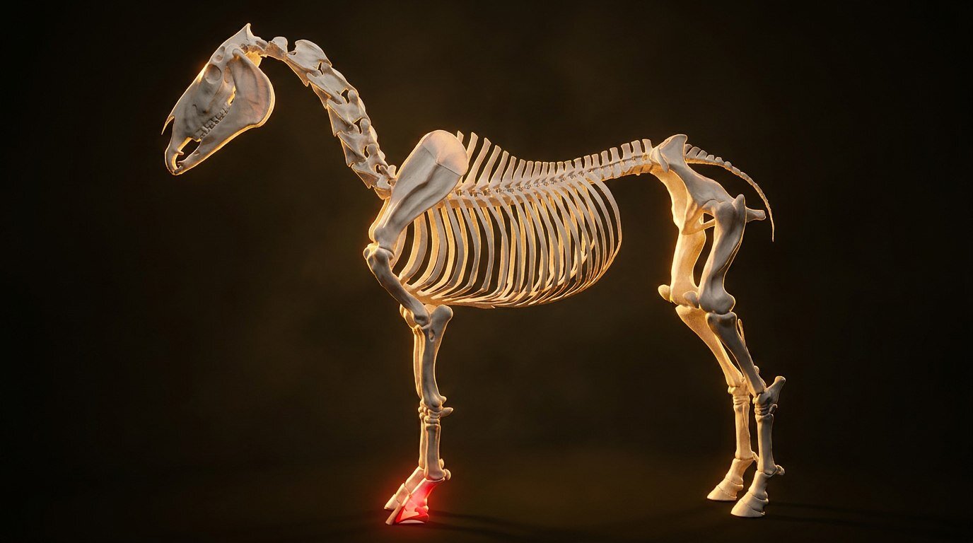
There is a particular kind of dread that settles in your gut when you walk out to the pasture and see your horse standing rock-still, leaning back on his heels like the ground in front of him is made of hot coals. You know what it might be. Every horse person does. Laminitis is one of the most common emergencies in equine medicine, and it has been destroying hooves and breaking hearts for as long as humans have kept horses. But here is the thing that frustrates me about how laminitis gets discussed: people throw the word around like it is one disease. It is not. Not even close.
What the Laminae Actually Are
Before you can understand what goes wrong, you need to understand what is in there. The hoof wall is not fused directly to bone. Between the inner surface of the hoof wall and the coffin bone (third phalanx, P3, distal phalanx, whatever name you prefer) sits an intricate structure called the laminae. Think of it like Velcro, except biological and load-bearing.
There are two sets. The epidermal laminae (also called insensitive laminae) project inward from the hoof wall. The dermal laminae (sensitive laminae) project outward from the coffin bone. These two sets interdigitate, folding into each other with secondary laminae branching off each primary lamina. The total surface area is staggering. Estimates put it around 800 square centimeters in an average-sized horse. All of that surface area exists for one purpose: suspending approximately 500 to 600 pounds of horse from inside the hoof capsule.
The basement membrane sits right at the junction between those epidermal and dermal laminae. It is a thin, specialized layer of extracellular matrix. When laminitis strikes, this is ground zero. The basement membrane degrades, the bond between the two laminar sets weakens, and the coffin bone starts to lose its suspension. For a deeper look at how the whole structure fits together, check out our hoof anatomy breakdown.
Three Roads to the Same Disaster
Veterinary science now recognizes three distinct pathways that lead to laminar failure. They share an endpoint but get there through completely different mechanisms.
Endocrine/Metabolic Laminitis
This is the big one. Roughly 90% of laminitis cases seen in clinical practice trace back to endocrine dysfunction. Two conditions dominate: Equine Metabolic Syndrome (EMS) and Pituitary Pars Intermedia Dysfunction (PPID, formerly called Cushing's disease).
The common thread is insulin. Horses with EMS have tissue insulin resistance paired with compensatory hyperinsulinemia. Their bodies pump out excessive insulin to manage blood glucose, and that chronically elevated insulin is directly toxic to the laminae. Researchers at the University of Queensland demonstrated this by infusing healthy horses with insulin while maintaining normal glucose levels. The horses developed laminitis. Insulin alone did it.
PPID works a bit differently. The pituitary gland's pars intermedia loses its dopaminergic inhibition, usually because of oxidative neurodegeneration, and starts overproducing ACTH and other pro-opiomelanocortin-derived peptides. The resulting hormonal chaos often produces insulin dysregulation as a downstream effect. Not every PPID horse is insulin-resistant, but plenty of them are, and those are the ones who founder.
The metabolic laminitis horse is the one you see most often: the easy keeper, the cresty-necked pony, the aging gelding with a curly coat who gets into the spring grass. That is not a coincidence. Pasture grasses accumulate fructans and simple sugars, especially during cool nights followed by sunny days. A metabolically compromised horse grazing those pastures is essentially mainlining a laminitis trigger.
Sepsis-Associated Laminitis
This type shows up in horses that are already fighting a systemic war. Grain overload, retained placenta, severe colic, pleuropneumonia, colitis. The common denominator is endotoxemia or a massive systemic inflammatory response. Bacterial endotoxins and inflammatory mediators circulate through the bloodstream and wreak havoc on the laminar vasculature and basement membrane.
The speed of septic laminitis is what makes it terrifying. A mare who retained her placenta can be showing laminar signs within 24 to 48 hours. The inflammatory cascade activates matrix metalloproteinases (MMPs), enzymes that chew through the basement membrane like acid through paper. By the time you see the horse shifting weight, significant structural damage may already be underway.
Mechanical/Supporting Limb Laminitis
This is the cruel irony of orthopedic injuries. A horse with a severe fracture, a septic joint, or post-surgical pain in one leg will load the opposite limb excessively. That contralateral hoof was never designed to bear 100% of the load for days or weeks on end. The sustained mechanical stress compromises blood flow to the laminae, and the tissue begins to fail.
Supporting limb laminitis is what killed Barbaro. His catastrophic breakdown at the Preakness led to surgical repair of the right hind, but the left hind developed progressive laminitis under the abnormal weight-bearing. It remains one of the most difficult forms to prevent because you cannot simply unload the supporting limb without addressing the primary injury first.
The Obel Grading System
Veterinarians commonly use the Obel scale to classify laminitis severity. It runs from Grade I through Grade IV.
Obel Grade I: The horse shifts weight from foot to foot at rest. Lameness is not obvious at the walk but becomes apparent at the trot, especially on turns or hard ground. You might notice the horse landing toe-first instead of heel-first.
Obel Grade II: The horse walks willingly but moves with a stiff, stilted gait. You can pick up a front foot, but the horse resists and quickly puts it back down. The classic "walking on eggshells" look starts here.
Obel Grade III: The horse is reluctant to move and resists attempts to lift a foot vigorously. The characteristic rocked-back stance is obvious, with the hind legs camped underneath the body to shift weight off the front end. Turning is painful. The digital pulse is usually bounding.
Obel Grade IV: The horse refuses to move. Period. May be recumbent. Forcing movement is cruel at this stage. These horses are in extreme pain and the prognosis is guarded to poor depending on radiographic findings.
Rotation, Sinking, and What Radiographs Tell You
Once the laminar bond fails, the coffin bone is no longer properly suspended. Two things can happen, and often both occur simultaneously.
Rotation means the tip of the coffin bone tilts downward, pivoting at the toe. The deep digital flexor tendon, which attaches to the palmar surface of P3, keeps pulling, and without the laminar counter-force, it wins. The bone rotates away from the hoof wall. On lateral radiographs, you measure the angle between the dorsal surface of the coffin bone and the dorsal hoof wall. Any divergence indicates rotation. More than 5.5 degrees is concerning. Double digits are serious.
Distal displacement, commonly called sinking, is worse. It means the coffin bone has dropped within the hoof capsule. The entire bone moves distally rather than just rotating. Sinking indicates failure across a broader area of laminae and carries a significantly worse prognosis. Severe sinking can result in the coffin bone penetrating through the sole.
Your vet will use the founder distance (the measurement between the proximal extent of the hoof wall and the extensor process of P3) to quantify sinking. Normal is roughly 0 to 5 mm depending on the horse. Values above 15 mm start getting into territory where long-term soundness becomes questionable.
Acute vs Chronic: The Timeline Matters
Acute laminitis is the inflammatory event itself, the active crisis where the laminae are under assault. This phase can last anywhere from a few days to a couple of weeks. During this window, the priority is pain management, removing the inciting cause, and supporting the hoof mechanically.
Chronic laminitis is what comes after. The laminar damage has occurred, the coffin bone has displaced to whatever degree it is going to, and the hoof begins to remodel. You will see divergent growth rings on the hoof wall (wider at the heels, tighter at the toe), a dished dorsal wall, a dropped or flat sole, and sometimes a stretched white line. A horse can be chronically laminitic without being actively lame, especially with good farrier care, but the structural changes are permanent.
The transitional period between acute and chronic, sometimes called the subacute phase, is tricky. The horse may seem to improve and then worsen. Radiographs taken too early can underestimate the final degree of displacement because the bone may still be moving. Serial radiographs over weeks give a much clearer picture.
Treatment in the Acute Phase
Speed matters. If you suspect laminitis, get your vet out immediately. Not tomorrow. Today.
Cryotherapy is one of the few interventions with solid evidence behind it. Cooling the feet to below 10 degrees Celsius (about 50 degrees Fahrenheit) reduces the enzymatic destruction of the basement membrane and limits the inflammatory cascade. The earlier you start, the better. Ideally, cryotherapy begins before clinical signs even appear in at-risk horses, such as a horse being treated for grain overload or a mare with a retained placenta. Ice boots, ice slurries in rubber boots, or simply standing the horse in a tub of ice water all work. Keep it going for 48 to 72 hours in high-risk cases.
NSAIDs provide pain relief and reduce inflammation. Phenylbutazone is the old standby. Flunixin meglumine (Banamine) gets used frequently as well. Some vets combine low-dose phenylbutazone with acetaminophen for multimodal analgesia. In severe cases, continuous rate infusions of lidocaine or other analgesics may be necessary.
Mechanical support for the hoof matters enormously. Styrofoam pads taped to the feet provide cheap, effective sole support. They compress under the horse's weight and distribute load across the frog and sole rather than concentrating it at the wall. Some farriers and vets prefer commercial frog support pads or impression material packed into the sole.
Stall rest on deep bedding is standard. Sand is ideal because it conforms to the hoof and provides consistent support. Shavings work too. The horse should not be forced to walk during the acute phase.
Long-Term Management and Rehab Shoeing
This is where good farriery becomes a lifeline. The goals of therapeutic shoeing in chronic laminitis are straightforward even if execution is complex: ease breakover, support the palmar/plantar structures, protect the sole, and realign the coffin bone with the hoof capsule over successive trim cycles.
Heart bar shoes were the traditional go-to. They apply direct pressure to the frog, providing some support to the coffin bone through the digital cushion. Modern approaches have expanded the toolkit considerably. Reverse shoes, rocker-toe shoes, and various cuff or rail systems all have their place. Many farriers now work with glue-on composite shoes for severely damaged feet because nailing into compromised hoof wall is asking for trouble.
What actually turns things around for many laminitic horses is the combination of proper trimming philosophy and radiographic guidance. The farrier trims to the coffin bone, using lateral radiographs taken with markers on the hoof to align trim angles with the bone's actual position. This is not a standard trim. It requires a farrier who understands laminitis mechanics and ideally works alongside a veterinarian.
For the metabolic laminitis horse, none of this matters if you do not address the underlying endocrine problem. That means restricted grazing, a low-sugar and low-starch diet (under 10% NSC), appropriate exercise as soundness allows, and possibly pergolide for PPID or metformin as an adjunct for EMS. You can explore the broader picture of hoof pathology at our laminitis condition page.
Regular monitoring is non-negotiable. Body condition scoring, insulin testing (ideally dynamic testing like the oral sugar test), and periodic radiographs to track hoof capsule changes should all be part of the plan. Too many horses founder again because their owners think the crisis is over once the lameness resolves. The metabolic time bomb is still ticking if you have not defused it.
The Emotional Side Nobody Talks About
Laminitis is relentless. It demands months of stall rest, meticulous diet management, expensive vet and farrier bills, and the constant anxiety of watching your horse for any sign of regression. Some horses recover beautifully. Others do not, and the conversation about humane endpoints has to happen honestly when suffering cannot be adequately managed. That is not a failure. That is responsible horse ownership at its hardest.
There is real hope, though. Advances in understanding the insulin-laminitis connection have given us better tools for prevention than we had even a decade ago. Horses that would have been lost are now being managed successfully with diet, drugs, and skilled farriery. Early recognition and early intervention change outcomes dramatically. Use our interactive models to study the hoof structures involved and build your understanding from the anatomy up.
Frequently Asked Questions
Can a horse fully recover from laminitis?
It depends on the severity. Horses with mild rotation (Obel Grade I or II) and prompt treatment often return to comfortable soundness and even light riding. Horses with significant sinking or severe rotation may remain pasture-sound at best. Full histological recovery of the laminae is unlikely once substantial damage has occurred, but functional recovery with careful management is achievable for many horses.
Is laminitis the same as founder?
Not exactly. Laminitis refers to the inflammation of the laminae. Founder specifically refers to the displacement of the coffin bone that results from laminar failure. So founder is a consequence of laminitis, but a horse can have laminitis that is caught and treated before founder (coffin bone displacement) occurs.
Why does grass cause laminitis?
Grass itself is not the enemy. The problem is the non-structural carbohydrates (sugars and fructans) that grasses accumulate, particularly during cool nights followed by warm sunny days, and during periods of plant stress. Horses with insulin dysregulation cannot process these sugars normally, leading to dangerous insulin spikes that damage the laminae. Healthy horses grazing the same pasture are usually fine.
How quickly does laminitis develop?
Septic laminitis from grain overload or retained placenta can develop within 24 to 48 hours. Endocrine laminitis from metabolic dysfunction tends to develop more gradually, sometimes over days to weeks of worsening subclinical damage before obvious lameness appears. Supporting limb laminitis can develop within days to weeks of abnormal weight-bearing.
Should I exercise a horse with chronic laminitis?
Only when the veterinarian and farrier both agree the horse is comfortable enough. Controlled exercise can actually benefit the metabolic laminitis horse by improving insulin sensitivity and promoting healthy hoof growth. But exercising a horse that is still actively lame accelerates damage. Let the professionals guide the timeline.
- Pollitt, C.C. "Equine Laminitis." Clinical Techniques in Equine Practice, 2004. University of Queensland.
- Asplin, K.E., Sillence, M.N., Pollitt, C.C., McGowan, C.M. "Induction of laminitis by prolonged hyperinsulinaemia in clinically normal ponies." The Veterinary Journal, 2007.
- American Association of Equine Practitioners (AAEP). "Laminitis: Prevention and Treatment." AAEP Guidelines.
- van Eps, A.W. "Therapeutic Hypothermia (Cryotherapy) to Prevent and Treat Acute Laminitis." Veterinary Clinics of North America: Equine Practice, 2010.
- Karikoski, N.P., Horn, I., McGowan, T.W., McGowan, C.M. "The prevalence of endocrinopathic laminitis among horses presented for laminitis at a first-opinion/referral equine hospital." Domestic Animal Endocrinology, 2011.
Sources
- Pollitt, C.C. "Equine Laminitis." Clinical Techniques in Equine Practice, 2004. University of Queensland.
- Asplin, K.E., Sillence, M.N., Pollitt, C.C., McGowan, C.M. "Induction of laminitis by prolonged hyperinsulinaemia in clinically normal ponies." The Veterinary Journal, 2007.
- American Association of Equine Practitioners (AAEP). "Laminitis: Prevention and Treatment." AAEP Guidelines.
- van Eps, A.W. "Therapeutic Hypothermia (Cryotherapy) to Prevent and Treat Acute Laminitis." Veterinary Clinics of North America: Equine Practice, 2010.
- Karikoski, N.P., Horn, I., McGowan, T.W., McGowan, C.M. "The prevalence of endocrinopathic laminitis among horses presented for laminitis at a first-opinion/referral equine hospital." Domestic Animal Endocrinology, 2011.
Want to prevent and manage laminitis effectively?
Our Lameness course covers founder, metabolic triggers, and rehabilitation protocols.
Explore the Lameness CoursePro members only. Try Pro free for 7 days.