Laminitis in Horses: Prevention, Signs, and Recovery
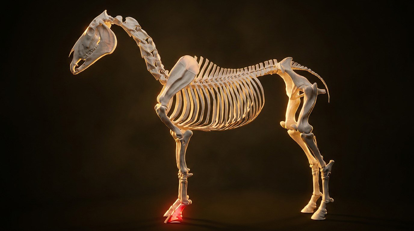
Laminitis in Horses: Prevention, Signs, and Recovery
A barn manager once told me she could set her watch by spring laminitis cases. First week of May, like clockwork, the phone starts ringing. Somebody's easy keeper got loose on the new grass. Somebody's pony looks "a little stiff." By the time the vet arrives, the damage has been brewing for 48 hours and the real fight is already behind them.
Laminitis in horses is one of those conditions where the gap between what owners think is happening and what's actually happening inside the hoof can be the difference between a horse that recovers and one that doesn't. The scariest part isn't the lameness you see. It's the structural destruction that starts before you see anything at all.
The Laminae: Why This Structure Matters So Much
To understand laminitis, you need to understand what the laminae actually do. Picture interlocking fingers: one set attached to the inside of the hoof wall (epidermal laminae) and the other attached to the coffin bone (dermal laminae). These two sets of tissue interlock in approximately 550-600 primary laminae, each with 100-200 secondary laminae. That's somewhere around 80,000 to 120,000 connection points per hoof.
This tissue suspends the entire weight of the horse inside the hoof capsule. The coffin bone doesn't sit on the sole like a foot in a shoe. It hangs from the hoof wall via the laminae, like a rock climber hanging from their fingers. When the laminae let go, the coffin bone has nothing holding it up.
You can explore the laminae and coffin bone inside the hoof capsule in our 3D model to see exactly how this suspension works. Rotating the view makes it immediately obvious why laminar failure is so catastrophic.
What Triggers Laminitis: The Three Pathways
Not all laminitis arrives the same way. The trigger matters because it shapes how you prevent it, how fast it progresses, and what the prognosis looks like.
Endocrine (Metabolic) Laminitis
This is the most common form. Equine Metabolic Syndrome (EMS) and Pituitary Pars Intermedia Dysfunction (PPID, formerly called Cushing's disease) are the two big players. Horses with EMS are insulin-dysregulated: their bodies overproduce insulin in response to sugars and starches, and chronically elevated insulin directly damages the laminae.
The classic EMS horse is the cresty-necked, overweight, easy keeper who gets fat on air. But don't be fooled by stereotypes. Lean horses can be insulin-dysregulated too, and PPID affects horses of any body type, usually over age 15. The patchy, curly coat that doesn't shed properly is the hallmark of PPID, but laminitis can appear before the coat changes do.
Endocrine laminitis often presents as chronic, low-grade, bilateral forelimb lameness that waxes and wanes with the seasons. Spring and fall are peak danger periods because pasture sugar content spikes during cool nights followed by sunny days.
Inflammatory (Sepsis-Related) Laminitis
Grain overload. Retained placenta. Severe colic. Pleuropneumonia. Any condition that triggers a systemic inflammatory response can cause acute laminar failure as a secondary complication. This is the form that terrifies equine veterinarians because it hits hard and fast.
Here's the mechanism that connects gut to hoof. When a horse gorges on grain, the sudden carbohydrate load overwhelms the small intestine's capacity to digest it. The undigested starch dumps into the cecum and hindgut, where it feeds lactic acid-producing bacteria. The gut pH plummets. Beneficial fiber-fermenting bacteria die off. The gut lining becomes permeable. Bacterial endotoxins leak into the bloodstream. Those circulating toxins trigger a cascade of inflammatory mediators that target the laminae.
The delay is what kills owners. A horse breaks into the feed room Saturday night. Sunday morning, he seems fine, maybe a little quiet. By Monday afternoon, he can barely walk. The grain hit the gut 36-48 hours ago, but the laminar damage took time to manifest. That 24-72 hour window between the insult and the clinical signs is when the laminae are actively being destroyed, and the owner often has no idea.
Mechanical (Supporting Limb) Laminitis
When a horse non-weight-bears on one leg due to a fracture, severe infection, or surgical recovery, the opposite limb bears excessive load. After days to weeks of this unrelenting weight, the laminae in the supporting limb begin to fail. This is how Barbaro was ultimately lost: not to the initial fracture, but to laminitis in the supporting limb.
Supporting limb laminar compromise is the most difficult form to prevent and carries the worst prognosis. Veterinary teams use frog support pads, controlled icing, and careful limb-loading protocols, but once it takes hold, options narrow quickly.
Three Types of Laminitis Compared
| Type | Trigger | Most Common In | Prevention Focus |
|---|---|---|---|
| Endocrine (Metabolic) | Chronically elevated insulin from EMS or PPID | Overweight, easy keepers, senior horses | Diet management, exercise, PPID testing |
| Inflammatory (Sepsis) | Grain overload, retained placenta, severe colic, systemic infection | Any horse after acute illness or dietary mishap | Secure feed rooms, prompt treatment of illness |
| Mechanical (Supporting Limb) | Prolonged weight-bearing on one limb due to contralateral injury | Horses with severe single-limb lameness | Aggressive support of the weight-bearing limb, frog support |
Early Signs: What 48 Hours Looks Like
Laminar inflammation has a timeline, and where you catch it on that timeline determines almost everything.
Developmental phase (0-24 hours post-trigger): The horse may appear normal. If you know the horse got into grain or has been on dangerously lush pasture, this is your window to act. Call your vet immediately. Don't wait for lameness. Ice the feet aggressively: continuous cryotherapy (ice boots or standing in ice water) for 48-72 hours has been shown to significantly reduce laminar damage when started during the developmental phase. The landmark Pollitt and van Eps research demonstrated that continuous digital hypothermia significantly reduces the severity of experimentally induced laminar damage.
Acute phase (24-72 hours): This is when you see it. The classic "founder stance," rocked back on the haunches with the front feet camped out ahead, trying to shift weight off the toes. Bounding digital pulses in the fetlock area. Heat in the hoof wall. Reluctance to walk, especially on hard surfaces. The horse may be visibly sweating, elevated heart rate, grinding teeth. This is an emergency.
Subacute/chronic phase: If the initial episode is survived and managed, the horse enters a recovery period that can last months. Radiographs taken 10-14 days post-onset reveal whether the coffin bone has rotated (the front of the bone tips downward) or sunk (the entire bone displaces distally). The degree of rotation or displacement directly predicts the difficulty of rehabilitation.
Emergency Response: What Your Vet Will Do
When your vet arrives for an acute laminar inflammation case, expect the following:
Pain management is immediate. NSAIDs (phenylbutazone, flunixin meglumine) reduce inflammation and pain. Severe cases may require additional analgesics. Pain control is not optional; an agonized horse that refuses to lie down or shift weight compounds the mechanical damage.
Cryotherapy if not already started. Continuous icing of the feet for 48-72 hours. This is not "put an ice pack on for 20 minutes." It means standing the horse in ice-water slurry or using commercial cryotherapy boots that maintain temperatures below 5°C at the hoof wall. The research on this is strong: icing works, but only if started early and maintained continuously.
Frog and sole support. Styrofoam pads cut to fit the sole, commercial frog support pads, or deep bedding redistribute load away from the compromised laminae and toward the frog, which can help bear weight as a secondary support structure.
Radiographs establish a baseline. Your vet measures the degree of rotation (normal is 0-2 degrees between the dorsal hoof wall and the dorsal surface of the coffin bone) and looks for displacement. Serial radiographs over the coming weeks track progression or stabilization.
Addressing the trigger. If the cause was grain overload, mineral oil via nasogastric tube to flush the gut. If the cause is endocrine, blood work to diagnose EMS or PPID so appropriate medication (pergolide for PPID, dietary management for EMS) can begin. If it's a retained placenta, that needs to come out. Treating laminitis without treating its cause is like mopping the floor while the faucet runs.
The Rehabilitation Road
Recovering from a founder episode is measured in months, not weeks. Hoof growth rate in horses averages about 6-10mm per month (our deep dive on hoof growth factors breaks this down), and a new, well-connected hoof capsule needs to grow from the coronary band all the way to the ground. That's 8-12 months minimum for a complete hoof capsule replacement.
During this time, your farrier becomes your horse's most important healthcare provider. Therapeutic trimming and shoeing for horses recovering from laminar compromise is specialized work. The farrier needs to:
- Back up the toe aggressively to ease breakover
- Maintain or restore appropriate palmar angle
- Apply frog support (pour-in pads, impression material, or heart bar shoes)
- Trim on a shortened cycle (every 3-4 weeks in the acute phase)
- Work from serial radiographs, not guesswork
| Phase | Focus | Details |
|---|---|---|
| Acute (Week 1–2) | Pain control & stabilization | Cryotherapy, NSAIDs, frog support, stall rest on deep bedding |
| Sub-acute (Week 2–8) | Hoof mechanics & monitoring | Therapeutic shoeing/trimming, serial radiographs, controlled diet |
| Recovery (Month 2–6+) | Gradual return to movement | Hand-walking, small paddock turnout, ongoing dietary management |
| Long-term | Prevention & maintenance | Regular trimming cycles, low-NSC diet, metabolic monitoring |
Movement during rehabilitation follows a careful progression. Stall rest with deep bedding initially, then hand-walking on soft ground, then small paddock turnout on non-grass surfaces. Returning to pasture requires testing the horse's insulin response and carefully managing grass exposure. A grazing muzzle is not cruelty; for an insulin-dysregulated horse, it might be the thing that prevents the next episode.
Prevention: The Stuff That Actually Works
Most laminar inflammation is preventable. That's the frustrating part. The same management mistakes cause the same crises, year after year.
Know your horse's metabolic status. A simple blood test (resting insulin and glucose, ideally an oral sugar test) tells your vet whether your horse is insulin-dysregulated. Test every horse over 10 years old, every easy keeper, every horse with a cresty neck. If you know your horse is metabolically challenged, you can manage for it. If you don't test, you're guessing.
Control pasture access. Strip grazing. Grazing muzzles. Dry lots with measured hay. Avoiding turnout on lush pasture during high-sugar periods (cool nights below 4°C followed by sunny days). Managing grass is less romantic than 24/7 turnout on rolling green fields, but for at-risk horses, unrestricted grazing is a loaded gun.
Secure your feed room. Every year, horses break into feed rooms and gorge on grain. Double-latch the door. Use horse-proof locks. This is one of the most preventable emergencies in equine medicine and one of the most common.
Maintain hoof health. Regular trimming and balanced feet reduce mechanical stress on the laminae. A long-toed, underrun-heel foot puts leverage on the laminar attachment with every step. Good routine hoof care isn't just cosmetic; it's structural maintenance. Read about laminitis as a condition for more clinical detail.
Treat PPID. Pergolide (Prascend) controls the hormonal dysfunction that drives PPID-related laminar compromise. It's inexpensive, well-tolerated, and effective. If your aging horse has an abnormal coat, regional fat deposits, or a history of laminitis, ask your vet to test ACTH levels.
Prognosis: Honest Expectations
Prognosis depends on the type, severity, and speed of intervention. Mild endocrine laminitis cases caught early, with less than 5 degrees of rotation and no sinking, carry a good prognosis. Many of these horses return to comfortable pasture life and light riding with dietary management and careful hoof care.
Moderate cases (5-10 degrees of rotation) face a longer rehabilitation but can often achieve pasture soundness. Severe cases (greater than 10 degrees of rotation, any degree of distal displacement, sole penetration by the coffin bone) carry a guarded to poor prognosis. The decision about humane euthanasia in severe laminitis is one of the hardest conversations in equine medicine, and it should be guided by a veterinarian who can honestly assess the horse's pain level and realistic outcomes.
The horses that recover best are the ones whose owners acted fast, iced early, and committed to months of careful hoof rehabilitation without cutting corners.
Frequently Asked Questions
Can a horse fully recover from laminitis?
Many horses recover from laminitis and return to comfortable lives, including light to moderate work. However, "fully recover" is nuanced. A horse that has foundered once is statistically more likely to founder again, especially if the underlying trigger (metabolic disease, pasture management) isn't addressed. The hoof may have permanent structural changes visible on radiographs even when the horse is clinically sound.
How quickly does laminitis develop?
The developmental phase can be as short as 8-12 hours (grain overload, sepsis) or as gradual as weeks to months (chronic endocrine laminitis). The inflammatory damage to the laminae typically precedes visible lameness by 24-72 hours. This is why acting on the trigger (rather than waiting for symptoms) is so critical.
Is laminitis always in the front feet?
Most commonly, yes. The front feet bear approximately 60% of the horse's weight, so they're more susceptible. However, laminitis can affect all four feet, particularly in severe endotoxemic cases. Hindlimb-only laminitis is uncommon but possible, especially with supporting limb laminitis.
Can you ride a horse that has had laminitis?
Potentially, depending on the severity and recovery. Horses that experienced mild laminitis with minimal coffin bone displacement and whose underlying metabolic issues are managed may return to light riding after 6-12 months of rehabilitation. Horses with significant rotation or chronic changes may be limited to pasture soundness. Your vet and farrier should make this call together based on serial radiographs and the horse's comfort level.
What's the difference between laminitis and founder?
Laminitis is the inflammation of the laminae. Founder specifically refers to the mechanical failure: when the coffin bone displaces (rotates or sinks) within the hoof capsule. All founder involves laminitis, but not all laminitis progresses to founder. Think of laminitis as the fire and founder as the structural collapse that follows if the fire isn't controlled.
How much does laminitis treatment cost?
Initial emergency care (vet visit, radiographs, pain management, cryotherapy supplies) typically runs $500-$1,500. Ongoing therapeutic farrier work every 3-5 weeks during rehabilitation adds $200-$400 per cycle. Blood work and medication for endocrine disease adds $50-$150 per month. A severe case requiring hospitalization can run $3,000-$10,000+. Budget realistically for 6-12 months of intensive management.
Sources
- Pollitt, C.C. (2004). Equine laminitis. Clinical Techniques in Equine Practice, 3(1), 34-44.
- van Eps, A.W., & Pollitt, C.C. (2006). Equine laminitis induced with oligofructose. Equine Veterinary Journal, 38(3), 203-208.
- van Eps, A.W., Pollitt, C.C., Leise, B.S., et al. (2012). Digital hypothermia inhibits early lamellar inflammatory signaling. Equine Veterinary Journal, 44(3), 241-249.
- Frank, N., & Tadros, E.M. (2014). Insulin dysregulation. Equine Veterinary Journal, 46(1), 103-112.
- Karikoski, N.P., Horn, I., McGowan, T.W., & McGowan, C.M. (2011). The prevalence of endocrinopathic laminitis among horses presented for laminitis at a first-opinion/referral equine hospital. Domestic Animal Endocrinology, 41(3), 111-117.
- Merck Veterinary Manual: Laminitis in Horses. Available at merckvetmanual.com.
- American Association of Equine Practitioners (AAEP). Laminitis Prevention and Treatment Guidelines.
Ready to learn the full spectrum of lameness conditions?
Our Lameness course covers laminitis, navicular, and every major cause of unsoundness.
Start the Lameness CoursePro members only. Try Pro free for 7 days.