The Stay Apparatus in Horses: How They Sleep Standing Up
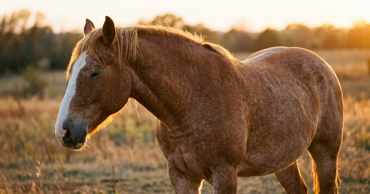
You have probably watched a horse doze in a field, one hind leg cocked, head drooping, completely unconscious to the world. Standing up. Asleep. On four legs. And you have probably wondered how on earth they do that without face-planting into the dirt. The answer is the stay apparatus, a brilliantly engineered system of tendons, ligaments, and locking mechanisms that allows a horse to support its body weight while expending almost zero muscular energy. It is one of the great feats of biological engineering in the animal kingdom.
Quick Answer: The stay apparatus is a system of ligaments, tendons, and anatomical locking mechanisms in the fore and hind limbs that allows horses to remain standing with minimal muscular effort. In the forelimb, a series of ligamentous and tendinous structures locks the shoulder, elbow, carpus, and fetlock in extension. In the hind limb, the reciprocal apparatus mechanically links the stifle and hock so they flex and extend together, while the patella physically locks over the medial femoral condyle to prevent the stifle from bending. This system evolved because prey animals on open plains needed to sleep while remaining ready to flee.
Why Standing Sleep Exists
The evolutionary logic is brutal and simple. Horses are prey animals that evolved on open grasslands where predators could appear without warning. A horse lying down needs several seconds to stand up. Several seconds is an eternity when a pack of wolves is closing in at 35 miles per hour. So horses evolved the ability to sleep, rest, and even achieve light sleep stages while standing, keeping all four legs underneath them and ready for instant flight.
Horses do still lie down. They have to. REM sleep, the deep dreaming stage, requires the complete muscle relaxation that only comes with recumbency. A horse that never lies down will eventually become sleep-deprived, which leads to a condition called narcolepsy-like collapse or sleep deprivation syndrome, where the horse buckles at the knees and drops without warning. Horses need about 30 minutes to 2 hours of recumbent sleep per day, typically taken in short bouts of 15 to 20 minutes. But for the other 22+ hours, they stand. And the stay apparatus makes that possible without exhaustion.
The Forelimb Stay Apparatus
The forelimb stay apparatus is more about passive tendinous support than active locking. It works by creating a column of support from the scapula to the hoof that requires minimal muscular effort to maintain.
Shoulder and Elbow
The shoulder joint (scapulohumeral joint) is stabilized primarily by the biceps brachii muscle and the lacertus fibrosus, a strong fibrous band that connects the biceps to the extensor carpi radialis muscle on the front of the forearm. When the horse is standing, the weight of the body falling behind the shoulder joint creates a tendency for the shoulder to flex. The biceps and lacertus fibrosus resist this passively through their tendinous structure without requiring active muscular contraction.
The elbow is locked in extension by the triceps tendon inserting on the olecranon (the point of the elbow). When the leg is bearing weight, the ground reaction force passes in front of the elbow joint, creating an extending moment that is resisted passively. The elbow essentially "locks" because of the geometry of the forces acting on it. No muscular effort needed.
Carpus (Knee)
The carpus locks in extension through a combination of joint surface geometry and ligamentous support. The back of the carpus has a prominent ridge on the radius and corresponding grooves in the carpal bones that prevent hyperextension. When the leg is loaded, the bones stack in a way that resists bending. The palmar carpal ligaments and the fibrous joint capsule hold everything in place.
Interestingly, horses cannot truly lock the carpus the way they lock the hind limb stifle. A sleeping horse does occasionally buckle at the knee, especially if startled or in particularly deep sleep. This is normal and does not indicate a problem. The forelimb system is less mechanically absolute than the hindlimb system.
Below the Knee: The Suspensory System
From the carpus down, the stay apparatus relies heavily on the suspensory ligament, the deep digital flexor tendon (DDFT), the superficial digital flexor tendon (SDFT), and the distal sesamoidean ligaments. Together, these structures prevent the fetlock from collapsing to the ground under the horse's weight.
The suspensory ligament (interosseous muscle, though it is almost entirely ligamentous in adult horses) originates from the back of the cannon bone and the bottom row of carpal bones. It runs down the back of the cannon bone, splits into two branches that attach to the sesamoid bones at the fetlock, and sends extensor branches forward to join the common digital extensor tendon. It is the primary support for the fetlock joint.
The SDFT and DDFT run down the back of the leg, pass over the sesamoid bones at the fetlock (using them as a pulley), and insert on the pastern bones and coffin bone respectively. They work with the suspensory ligament to support the fetlock and prevent hyperextension. The superior check ligament (accessory ligament of the SDFT) and the inferior check ligament (accessory ligament of the DDFT) connect these tendons to the bones of the forearm and cannon, converting them into passive, ligament-like structures during standing.
The check ligaments are the secret weapons of the forelimb stay apparatus. By anchoring the flexor tendons to bone, they transfer the load from the muscles (which would require energy to contract) to the ligaments (which hold passively without energy expenditure). A horse standing on three legs while resting the fourth is supporting hundreds of pounds per limb through structures that burn essentially no calories to do their job.
The Hind Limb Stay Apparatus
The hind limb system is more complex and more mechanically complete than the forelimb system. It includes two components that work together: the reciprocal apparatus and the patellar locking mechanism.
The Reciprocal Apparatus
This is a mechanical linkage system that forces the stifle and hock to flex and extend together. You cannot bend one without bending the other. Try it on a cadaver limb and you will find it physically impossible to flex the stifle while holding the hock in extension, or vice versa.
The linkage is created by two structures:
- Peroneus tertius (fibularis tertius): A completely tendinous structure (no muscle belly at all in horses) that runs from the front of the femur, over the front of the tibia, to the front of the hock. When the stifle flexes, the peroneus tertius pulls the hock into flexion. If this structure ruptures, which is uncommon but does happen, the horse can extend the hock while the stifle is flexed, creating a bizarre overextension of the hock that is immediately recognizable.
- Superficial digital flexor tendon (hind limb): In the hind limb, the SDFT has a strong tendinous component that runs from the back of the femur, over the point of the hock (calcaneus), and down to the digit. It prevents the hock from flexing while the stifle is extended. If it ruptures, the hock flexes excessively while the stifle stays extended.
Together, these two structures create a rigid parallelogram. The femur and tibia are two sides, the peroneus tertius is the front connecting rod, and the SDFT is the rear connecting rod. The hock and stifle must move in concert. This saves enormous energy because the horse only needs to control one joint, and the other follows automatically.
The Patellar Locking Mechanism
This is the most dramatic part of the stay apparatus. The horse can physically lock its stifle joint in extension by hooking the patella (kneecap) over the medial (inner) condyle of the femur.
The equine stifle has three patellar ligaments: lateral, middle, and medial. In other species, there is typically one. The medial patellar ligament hooks over the medial trochlear ridge of the femur, and the patella rotates slightly to catch on this ridge. Once locked, the stifle cannot flex, which means (thanks to the reciprocal apparatus) the hock cannot flex either. The entire hind limb becomes a rigid column from hip to hoof.
To unlock, the horse contracts the quadriceps muscles briefly to lift the patella up and over the ridge, releasing the lock. This is a quick, almost imperceptible movement in a healthy horse. You might see a slight jerk or catch as the horse initiates movement from rest, but it should be smooth and effortless.
When Locking Goes Wrong: Upward Fixation of the Patella
When the patellar locking mechanism fails to release properly, the patella gets stuck in the locked position. This is called upward fixation of the patella, and it is one of the most common orthopedic problems in horses, particularly young, unfit, or straight-legged individuals.
Mild cases show as an intermittent catching or clicking of the hind leg, especially at the walk or when transitioning from halt to walk. The leg may momentarily lock in extension and then release with a visible jerk. Moderate cases involve the leg locking for several strides before releasing. Severe cases result in the leg locking completely, dragged behind the horse in rigid extension until manually unlocked (by backing the horse or pushing the patella medially and upward).
Treatment depends on severity. Mild to moderate cases often respond to a conditioning program that builds the quadriceps muscles, particularly through hill work, trot work, and backing exercises. Stronger quadriceps provide more efficient release of the locking mechanism. Corrective farriery (sometimes a small lateral heel wedge or toe rocker) can help by altering hoof breakover and changing the forces at the stifle. Severe or refractory cases may require medial patellar ligament desmotomy (cutting the medial patellar ligament), though this surgery is somewhat controversial because it eliminates the locking ability entirely and may predispose to other stifle problems.
Below the Hock: Mirroring the Forelimb
The structures below the hock in the hind limb are essentially the same as those below the knee in the forelimb: suspensory ligament, SDFT, DDFT, check ligaments, and sesamoid bones all functioning to support the fetlock passively. The hind suspensory ligament has a slightly higher proportion of muscle tissue than the fore, but the principle is identical. These structures create a passive support column from hock to hoof that requires no muscular energy to maintain.
The Stay Apparatus and Horse Management
Fitness and the Stay Apparatus
The patellar locking mechanism relies on adequate quadriceps strength and proper limb conformation. Horses that are out of shape, growing rapidly, or have very straight hind leg conformation are more prone to locking issues. A progressive conditioning program is both preventive and therapeutic. Hill work is particularly valuable because walking uphill requires quadriceps engagement and strengthens the exact muscles that control patellar release.
Conformation Considerations
Horses with very straight stifle angles ("post-legged" conformation) are predisposed to upward patellar fixation because the straighter angle makes it easier for the patella to hook over the ridge and harder for it to release. Conversely, horses with very angled ("sickle-hocked") hind legs place more stress on the reciprocal apparatus and the structures below the hock. Neither extreme is ideal. Moderate angulation provides the best balance between efficient locking and easy release.
Hoof Care Connection
Long toes and low heels in the hind feet delay hoof breakover, which keeps the stifle in extension longer during each stride and increases the tendency for patellar locking. Proper trimming and shoeing that promotes a timely breakover can significantly reduce locking episodes. A squared or rolled toe is a common farriery approach for horses with mild locking issues.
Sleep Deprivation
Because the stay apparatus enables standing rest, it is easy to assume that a horse that stands all the time is sleeping fine. This is not necessarily true. Horses need to lie down for REM sleep, and some horses are prevented from lying down by social dynamics (low-ranking horse in a herd with limited safe space), physical problems (arthritis making it painful to get up), or environmental factors (wet, muddy, or too-small enclosures). A horse that collapses suddenly to its knees, sometimes scraping its fetlocks raw in the process, may be experiencing sleep deprivation episodes rather than neurological disease. Ensuring adequate safe lying space is an often-overlooked aspect of horse management.
Comparative Anatomy
Other large herbivores have similar but less sophisticated systems. Cattle can lock their stifles but less efficiently than horses. Elephants have pillar-like legs with very little flexion at rest, essentially using skeletal stacking rather than ligamentous locking. Giraffes, interestingly, almost never lie down and have a stay apparatus that is even more extreme than the horse's. Among domestic animals, the horse's stay apparatus is probably the most refined and energy-efficient system for sustained standing.
📝 Explore more in our Courses. Check it out here.
Frequently Asked Questions
Do horses actually sleep standing up or just rest?
Both. Horses achieve light sleep (slow-wave sleep) while standing, using the stay apparatus to support their weight. They cycle through drowsiness and light sleep throughout the day and night. However, they cannot reach REM sleep while standing because REM involves complete muscle relaxation, which would disable the active components of the stay apparatus and cause the horse to collapse. Horses must lie down for 30 minutes to 2 hours daily to get adequate REM sleep, usually in several short bouts.
Why does my horse's hind leg "catch" when it starts walking?
That catching sensation is likely the patella releasing from its locked position on the medial femoral condyle. In mild cases, this is a normal part of the unlocking process. If it is pronounced, frequent, or accompanied by a visible jerk or the leg locking in extension for multiple strides, it may indicate upward fixation of the patella. Young, unfit, or straight-legged horses are most commonly affected. Conditioning programs focusing on quadriceps strengthening (hill work, trot work, backing) usually improve mild cases. Persistent or severe cases should be evaluated by a veterinarian.
Can the stay apparatus be damaged?
Individual components can be injured. Rupture of the peroneus tertius, though uncommon, results in the ability to extend the hock while the stifle is flexed, which is immediately obvious. Suspensory ligament injuries are common athletic injuries that compromise the passive support of the fetlock. Check ligament injuries affect the ability of the flexor tendons to function as passive support structures. Complete failure of the stay apparatus as a whole does not really occur because it is made up of many independent structures, but damage to key components can significantly impair a horse's ability to rest comfortably while standing.
Is it bad if a horse never lies down?
Yes. A horse that never lies down will become REM-sleep deprived, which eventually causes involuntary collapse episodes. The horse may buckle at the knees, drop to the ground suddenly, or sway and stumble before catching itself. Over time, these episodes can cause injuries to the knees, fetlocks, and face. If your horse never lies down, investigate why. Common reasons include insufficient space, unsafe footing, social pressure from herdmates, pain that makes rising difficult (arthritis, laminitis), and anxiety in a new environment. Providing adequate safe space with comfortable, dry footing is essential.
Do foals have a functional stay apparatus?
Foals are born with the basic anatomical structures, but the stay apparatus is not fully functional in the first days of life. Newborn foals spend a much higher proportion of time lying down than adult horses, partly because their ligamentous structures are still lax and partly because they need more REM sleep for brain development. As the foal grows and the ligaments and tendons tighten, the stay apparatus becomes increasingly effective. By a few weeks of age, foals can stand and doze like adults, though they still lie down more frequently than mature horses.
- Budras, Klaus-Dieter, Sack, W.O., and Rock, Sabine. Anatomy of the Horse, 6th Edition. Schluetersche, 2012.
- Dyson, Sue. "The Stifle." Diagnosis and Management of Lameness in the Horse, 2nd Edition. Elsevier, 2011.
- Clayton, Hilary M. The Dynamic Horse. Sport Horse Publications, 2004.
- Houpt, Katherine A. "Sleep and Sleep Disorders in Horses." Equine Veterinary Education, 2020.
Last reviewed: June 2026
Sources
- Budras, Klaus-Dieter, Sack, W.O., and Rock, Sabine. Anatomy of the Horse, 6th Edition. Schluetersche, 2012.
- Dyson, Sue. "The Stifle." Diagnosis and Management of Lameness in the Horse, 2nd Edition. Elsevier, 2011.
- Clayton, Hilary M. The Dynamic Horse. Sport Horse Publications, 2004.
- Houpt, Katherine A. "Sleep and Sleep Disorders in Horses." Equine Veterinary Education, 2020.
Last reviewed: June 2026
Fascinated by equine biomechanics?
Our Anatomy course explains the stay apparatus and every structural adaptation.
Explore the Anatomy CoursePro members only. Try Pro free for 7 days.