Tendons vs Ligaments in the Horse: Structure, Injury, and Recovery
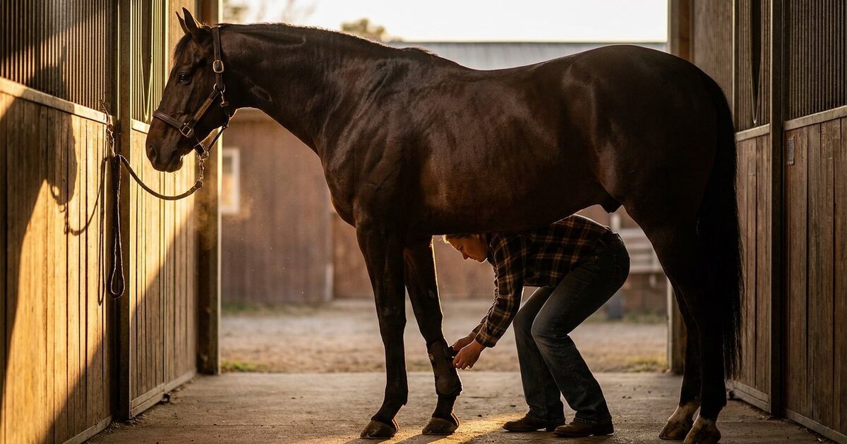
Your horse comes in from the pasture with a swollen leg. The vet says "soft tissue injury" and suddenly you're drowning in terms like SDFT, DDFT, suspensory, check ligament, and ultrasound grades. Tendons and ligaments are the unsung heroes of equine locomotion, and when they fail, the consequences range from frustrating to career-ending. Knowing the difference between these structures, and understanding how they heal, puts you in a much better position to make smart management decisions.
The Basics: What Connects to What
Tendons: Muscle to Bone
A tendon is the rope-like structure that connects a muscle belly to a bone. When the muscle contracts, the tendon transmits that force to move the skeleton. Simple enough. But in the horse's lower leg, tendons do something extra. They store and release elastic energy like biological springs.
The horse's lower leg has no muscles below the knee (carpus) and hock (tarsus). Everything down there is tendons, ligaments, and bone. This is an evolutionary masterpiece of energy efficiency. When a horse's hoof hits the ground at a gallop, the superficial digital flexor tendon (SDFT) stretches up to 12-16% of its resting length, storing kinetic energy that gets released during push-off. This saves roughly 36% of the metabolic energy that would otherwise be needed for locomotion.
That's incredible engineering. It's also why the SDFT is the most commonly injured tendon in sport horses. You're asking a biological tissue to repeatedly stretch to near its failure point, thousands of times per workout.
Ligaments: Bone to Bone
Ligaments connect one bone to another and primarily function to stabilize joints. They limit excessive range of motion and prevent bones from moving in directions they shouldn't. Unlike tendons, ligaments aren't meant to stretch much. They're designed to be stiff and restrictive.
The horse's suspensory ligament is a unique structure. It's technically a modified muscle (the interosseous muscle) that has largely lost its muscle fibers through evolution, becoming predominantly ligamentous. It runs down the back of the cannon bone, splits into two branches, and attaches to the sesamoid bones. It's critical to supporting the fetlock joint during weight bearing.
Structural Differences Under the Microscope
Both tendons and ligaments are primarily composed of Type I collagen fibers. But the organization is different.
Tendons have highly organized, parallel collagen fibers. Think of a steel cable: all the wires run in the same direction to maximize tensile strength along one axis. This parallel arrangement gives tendons enormous strength when pulled lengthwise but makes them vulnerable to shear forces from the side. The SDFT can withstand loads exceeding 12,000 Newtons in a 500 kg horse.
Ligaments have a more irregular, interwoven collagen arrangement. The fibers run in multiple directions, giving ligaments the ability to resist forces from various angles. This makes sense given that joints experience multi-directional stress. Ligaments also contain more elastin than tendons (roughly 5-15% vs. 1-2%), giving them slightly more inherent flexibility.
Blood Supply: Why It Matters for Healing
Blood supply is the critical factor in healing time, and this is where things get sobering. Tendons have notoriously poor blood supply, especially in their mid-body regions. The SDFT in the mid-metacarpal area receives blood primarily from its outer sheath (the paratenon), and the central core of the tendon is relatively avascular. This is the exact zone where most injuries occur.
Ligaments generally have slightly better blood supply than tendons, but it's still limited compared to muscle or skin. The suspensory ligament has a somewhat better vascular network than the flexor tendons, which partially explains why suspensory injuries, while serious, sometimes heal more reliably than SDFT tears.
Common Injuries: What Goes Wrong and Why
Bowed Tendon (SDFT Injury)
The classic "bowed tendon" is an injury to the superficial digital flexor tendon. It's called a "bow" because the swelling creates a visible outward curve on the back of the cannon bone region. This is the injury that strikes fear into racehorse trainers and eventers alike.
SDFT injuries typically happen during high-speed work or landing from jumps, when the tendon is loaded beyond its failure threshold. Fatigue is a major factor. A fresh tendon at the start of a workout can handle loads that a fatigued tendon 30 minutes later cannot. Research from the Royal Veterinary College in London found that cumulative microdamage from repeated loading weakens tendon fibers over time, eventually reaching a tipping point where clinical injury occurs.
The incidence in Thoroughbred racehorses is roughly 8-12% during racing careers. In eventers, it's estimated at 4-8% over a competitive lifetime. These are not small numbers.
Deep Digital Flexor Tendon (DDFT) Injury
DDFT injuries are less common in the mid-cannon region but increasingly recognized within the hoof capsule, where the tendon wraps around the navicular bone and inserts on the coffin bone. These injuries are closely associated with navicular syndrome and are a major cause of forelimb lameness in middle-aged sport horses. Diagnosis requires MRI, as ultrasound cannot image structures inside the hoof.
Suspensory Ligament Injury
Suspensory injuries come in three flavors: proximal (top), body (middle), and branch (bottom, where it splits). Each has different implications.
Proximal suspensory desmitis (PSD) in the hind limbs has become almost epidemic in dressage horses. Some estimates suggest it affects up to 15% of high-level dressage horses at some point. The repetitive collection work loads the proximal suspensory heavily, and the confined anatomy at the top of the cannon bone makes this area vulnerable.
Branch injuries are common in racehorses and show jumpers and generally carry a better prognosis than body or proximal injuries, with return-to-work rates around 60-70%.
Check Ligament Injuries
The inferior check ligament (accessory ligament of the DDFT) connects the cannon bone to the deep digital flexor tendon. It acts as a mechanical stop, preventing the DDFT from over-stretching. Injuries to this structure were once considered career-ending but respond reasonably well to controlled rehabilitation, with return-to-work rates around 75-80%.
Diagnosis: Ultrasound and Beyond
Ultrasound is the primary diagnostic tool for tendon and ligament injuries in the horse's leg. A skilled ultrasonographer can identify fiber disruption, measure lesion size, characterize the injury (acute inflammation vs. chronic scar tissue), and track healing over time.
Key ultrasound findings include cross-sectional area enlargement (a damaged tendon swells), hypoechoic (dark) lesions indicating fiber disruption, and loss of the normal parallel fiber pattern on longitudinal views. A normal SDFT in the mid-metacarpal region measures roughly 10-12 mm in diameter. An acutely injured SDFT might measure 15-20 mm.
MRI is used when ultrasound can't reach (inside the hoof) or when the diagnosis is unclear. Standing MRI units have made this much more accessible in the last decade, though a scan still runs $1,500-3,000 at most referral hospitals.
Healing: Why It Takes So Long
Tendon and ligament healing follows three phases:
Inflammatory phase (0-2 weeks): The body sends inflammatory cells to clean up damaged tissue. The leg is hot, swollen, and painful. This phase is actually necessary and productive, even though it looks alarming.
Proliferative phase (2 weeks - 4 months): The body lays down Type III collagen (scar tissue) to fill the defect. This new tissue is disorganized and weaker than the original tendon. It's functional but not yet mature.
Remodeling phase (4-18 months): Gradually, the Type III collagen is replaced by Type I collagen, and the fibers begin to align in the direction of stress. This is where controlled exercise becomes critical. The tendon needs mechanical loading to organize its new fibers properly. Too much load too soon causes re-injury. Too little load produces weak, disorganized scar tissue.
Here's the hard truth: healed tendon tissue is never as strong or elastic as the original. Studies show that repaired tendon has roughly 70-80% of the original tissue's mechanical properties, even after complete clinical healing. This is why re-injury rates for SDFT lesions run 27-42% depending on the study and the horse's intended use.
Rehabilitation: The Controlled Exercise Program
Modern tendon rehabilitation follows a graduated exercise protocol, typically spanning 9-18 months. The general framework:
Months 1-2: Stall rest with hand walking, starting at 10 minutes and building to 30-45 minutes daily. Ultrasound monitoring at 60 and 90 days.
Months 3-4: Increasing walk duration. Small paddock turnout may begin if the horse is sensible. Walking under saddle by month 4 for many protocols.
Months 5-8: Introduction of trot work, starting at 2-3 minutes and slowly increasing. This is the highest-risk phase for re-injury because riders get impatient and push too fast.
Months 9-12: Gradual return to canter work and discipline-specific training. Ultrasound monitoring continues every 8-12 weeks.
Months 12-18: Full return to work if ultrasound shows mature tissue and the horse remains sound.
Every horse is different. Some heal faster. Some slower. The ultrasound dictates the timeline, not the calendar. Your vet will tell you when to progress. Listen to them, not to your desire to ride.
Emerging Treatments
Several biological therapies have shown promise for improving tendon healing quality:
PRP (Platelet-Rich Plasma): Injecting concentrated platelets from the horse's own blood into the lesion. Growth factors in platelets stimulate healing. Evidence is mixed but generally favorable for acute injuries.
Stem Cell Therapy: Mesenchymal stem cells harvested from bone marrow or adipose tissue, then injected into the lesion. Multiple studies from Cornell and the Royal Veterinary College show improved tissue quality and reduced re-injury rates (roughly 18% vs. 42% for conventional treatment in one RVC study).
Shockwave Therapy: Extracorporeal shockwave can stimulate blood flow and healing in chronic lesions. It's more commonly used for ligament injuries than acute tendon tears.
None of these are magic bullets. They're adjuncts to controlled rehabilitation, not replacements for it. The boring part (the 12 months of walking) is still the foundation of tendon healing.
Frequently Asked Questions
Can a horse fully recover from a bowed tendon?
Many horses return to work after SDFT injuries, but "fully recover" depends on your definition. The tissue heals, the horse becomes sound, and many return to athletic careers. However, the repaired tissue is structurally different from the original, and re-injury rates are significant (27-42%). Horses returning to lower-level work have better long-term outcomes than those returning to racing or upper-level eventing.
How can I prevent tendon injuries?
Proper conditioning is the single best prevention. Gradual increases in workload. Adequate warm-up before intense exercise. Appropriate footing (deep or hard footing increases tendon strain). Monitoring leg temperature and swelling after workouts. Tendons adapt to training loads, but they adapt more slowly than muscle or cardiovascular fitness. Build your horse's tendon conditioning over months, not weeks.
What's the difference between tendonitis and desmitis?
Tendonitis is inflammation or injury to a tendon. Desmitis is inflammation or injury to a ligament. The "-itis" suffix means inflammation. You'll hear "suspensory desmitis" (ligament injury) and "superficial digital flexor tendonitis" (tendon injury). Same type of problem in different structures.
Are hind limb tendon injuries different from forelimb?
Yes. The forelimbs carry approximately 60% of the horse's body weight and bear the brunt of concussive force, making SDFT injuries primarily a front-leg problem. Hind limbs are more prone to suspensory ligament injuries, particularly proximal suspensory desmitis, due to the propulsive forces generated by the hindquarters. Treatment principles are similar, but the biomechanical causes differ.
When should I call the vet for a swollen leg?
Any acute swelling with heat and lameness warrants a vet call within 24 hours. If the horse is non-weight-bearing or the swelling is severe, call immediately. Early diagnosis and treatment (including icing and anti-inflammatories in the acute phase) significantly improve outcomes. Don't "wait and see" with potential tendon or ligament injuries. The first 48 hours of management matter enormously.
📝 Explore more in our Courses. Check it out here.
Sources
- Dyson, S. (2004). Medical management of superficial digital flexor tendonitis: a comparative study in 219 horses. Equine Veterinary Journal, 36(5), 415-419.
- Smith, R.K.W., et al. (2003). Clinical significance of tendon and ligament injuries. Royal Veterinary College, University of London.
- Merck Veterinary Manual. Tendon Disorders in Horses. merckvetmanual.com
- AAEP (American Association of Equine Practitioners). Lameness and Soft Tissue Injury Guidelines. aaep.org
- Goodship, A.E., Birch, H.L., & Wilson, A.M. (2012). The pathobiology and repair of tendon and ligament injury. Veterinary Clinics: Equine Practice, 10(2), 323-349.
Last reviewed: April 2026