Why Do Horses Have Hooves Instead of Toes?
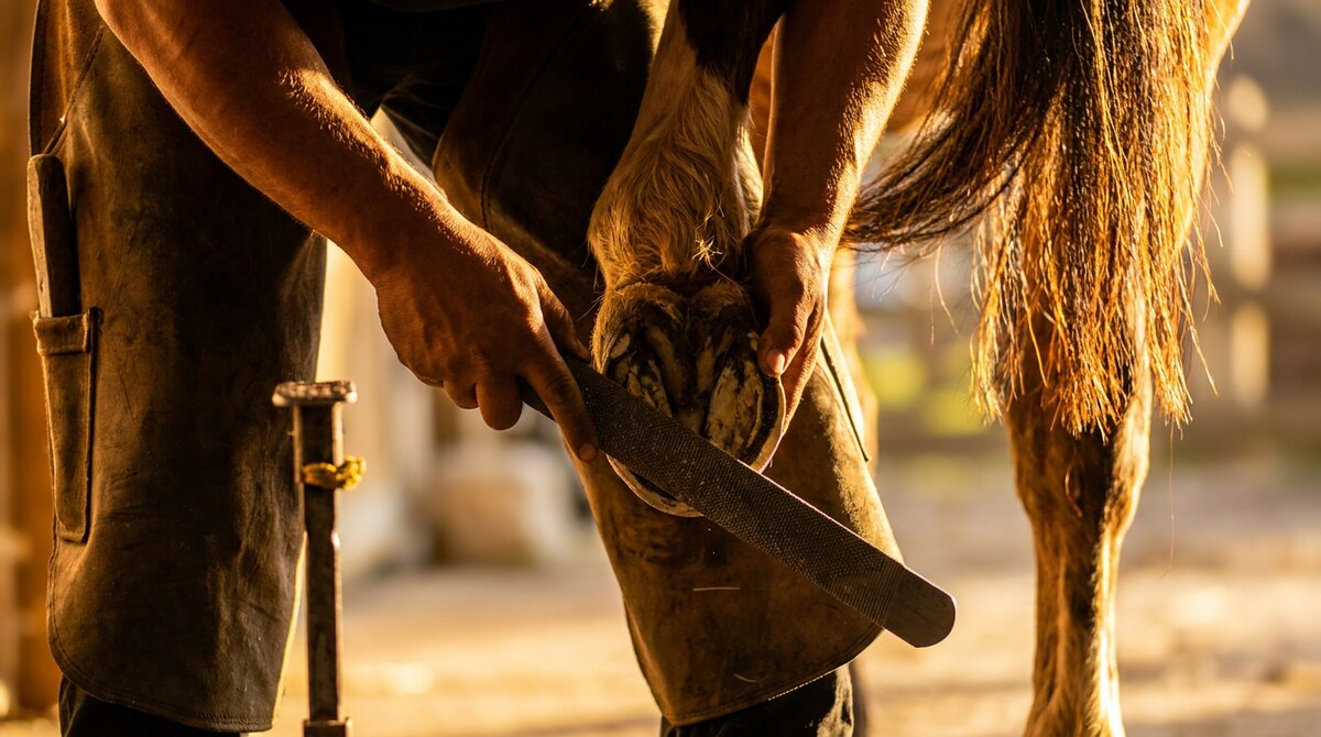
A horse gallops across a field on the tips of four middle fingers. That is not a metaphor. The hoof is a single toe, the anatomical equivalent of your middle finger, and the horse walks on the very tip of it, on a nail that has grown thick and hard enough to carry half a ton at speed.
How a four-toed, dog-sized forest animal became a one-toed runner is one of the best-documented stories in evolution, and the evidence is still visible in every horse's leg today.
The Horse Used to Have More Toes
The earliest horse ancestors looked nothing like a horse. The animal often called Eohippus (also known as Hyracotherium), living around 55 million years ago, was about the size of a small dog. It had four toes on each front foot and three on each hind foot, each toe with its own small hoof and a soft pad underneath, much like a modern tapir. It lived in forests and ate soft leaves and fruit, and its spread, padded feet were well suited to soft, uneven forest floor.
Then the world changed. Over millions of years, climate shifts turned many forests into open grassland. And open grassland is a completely different survival problem.
Why Grassland Favors One Toe
On open ground, there is nowhere to hide. A prey animal's best defense becomes speed and endurance, the ability to see a predator coming and outrun it over distance.
Long, light legs run faster and tire less. And the lightest, most efficient leg is one built from few bones, with as little weight as possible down at the far end where it swings fastest. A foot with multiple spreading toes is heavier and less efficient for sustained running than a single strong column ending in one toe.
So over the generations, natural selection did what it does. Horses with a larger, stronger central toe and reduced side toes ran better, survived better, and bred. The middle toe grew dominant. The side toes bore less and less weight and gradually shrank.
The fossil record tracks this in steps. Mesohippus, around 35 million years ago, had three toes with the middle one enlarging. Merychippus, around 10 million years ago, looked much more horse-like, still with three toes but with the central one bearing nearly all the weight and the side toes lifting off the ground. And finally Equus, the modern horse genus, emerged around 5 million years ago, standing on a single toe.
The Toes Are Still There
Here is the part that makes this real rather than just a story about the distant past. The lost toes did not vanish. They are still in the leg.
Run your hand down the cannon bone of any horse and you will feel, along each side, a thin bone tapering to a point partway down. Those are the splint bones. They are the reduced remnants of the second and fourth foot bones, the leftovers of two of the toes the horse's ancestors used to have. Every horse carries them. When a horse gets "splints," it is inflammation involving these evolutionary leftovers.
The evidence goes even deeper. Research has shown that early horse embryos briefly develop the beginnings of all five digits before most of them are reduced away, echoing the ancient five-toed pattern. And some studies have found faint ridges on the modern horse's foot bones interpreted as vestiges of the other lost digits. The horse's single toe is haunted by the ghosts of the ones it gave up.
You can see the single main digit and the two splint bones clearly in the 3D explorer with the skeletal layer on the lower limb. Once you have seen it, you cannot unsee that a horse is running on one finger.
The Hoof Is a Giant Toenail
The hoof wall, the hard outer part you nail a shoe to, is made of keratin, the same protein as your fingernail and your hair. It grows continuously down from the coronary band at the top, just as your nail grows out from the base, and it wears away at the bottom through contact with the ground. A horse that is not worn down naturally needs trimming for the same reason you trimming schedule your nails: the growth never stops.
Inside that keratin wall sits the coffin bone, the actual bone of the toe, suspended from the wall by the interlocking hoof anatomy. The whole structure is a single toe that has been reinforced, thickened, and turned into a running and shock-absorbing tool. The hoof anatomy in detail shows how the parts fit together.
Why It Matters
This is not only a good story. It explains real things about living with horses. It explains why splints exist and where they are. It explains why the hoof grows forever and needs a farrier. It explains why a horse's leg has no muscle below the knee, because the whole limb has been reduced to the lightest possible running column. And it explains why hoof and lower-limb problems are so central to horse health: the horse bet everything on one toe per leg, and when that toe has a problem, the horse has a problem.
"No hoof, no horse" is an old saying, and evolution is the reason it is true.
A horse's hoof is a single toe. Horses evolved from small, multi-toed forest ancestors over about 55 million years. As forests became open grassland, natural selection favored long, light legs and a single strong central toe for fast, efficient running, so the side toes shrank away and the middle toe became the hoof.
How many toes did horses used to have? The early horse ancestor Eohippus, around 55 million years ago, had four toes on each front foot and three on each hind foot. Over millions of years the number reduced, through three-toed forms, to the single toe of the modern horse.
What are splint bones? The splint bones are two thin bones running along the sides of a horse's cannon bone. They are the reduced remnants of two of the toes the horse's ancestors had, specifically the second and fourth foot bones. Every horse still carries them.
Is a horse's hoof really a toenail? The hoof wall is made of keratin, the same material as a human fingernail, and it grows continuously from the top and wears away at the bottom just as a nail does. The hoof is best understood as a single, greatly enlarged and thickened toe with a nail-like wall.
What is the horse's single toe called? It is the third digit. In the forelimb the bones are the third metacarpal (the cannon bone) and the phalanges below it, ending in the coffin bone inside the hoof. It corresponds to the middle finger of a human hand.
55 Million Years of Hoof Evolution
The story starts with Eohippus (Hyracotherium), a fox-sized forest dweller that walked on four toes on the front feet and three on the back. As grasslands replaced forests across North America roughly 35 million years ago, equine ancestors that could run faster survived. Over millions of years, the side toes shortened and the central toe strengthened, eventually producing the single-toed coffin bone we see today.
The vestigial splint bones on either side of the cannon bone are the remnants of those lost toes. Occasionally a horse is born with small extra digits alongside the hoof, a condition called atavism or polydactyly. These throwbacks to ancient anatomy are rare but well-documented.
The Hoof as an Engineering Marvel
A horse's hoof is not just a big toenail. It is a complex organ that manages forces of 2 to 3 times the horse's body weight at every stride. The hoof wall is made of densely packed tubular keratin that resists compression and wear. The sole protects the bottom, while the frog acts as a shock absorber and circulatory pump.
Inside, the laminae create an interlocking bond between the hoof wall and the coffin bone. There are roughly 600 primary epidermal laminae, each with 100 to 200 secondary laminae. This gives an estimated total surface area of attachment around 8 square feet per hoof. When this attachment fails, as in laminitis, the coffin bone can rotate or sink within the hoof capsule.
Blood Flow and the Hoof Mechanism
The digital cushion sits above the frog and is packed with blood vessels, fat, and fibrocartilage. When the hoof hits the ground, the frog compresses the digital cushion, which squeezes blood upward through the venous plexus. This mechanism supplements the heart's pumping action and is why movement is critical for hoof health.
Horses that stand in stalls all day develop poorer circulation in their feet. The coronary band, which sits at the top of the hoof capsule, is the growth center that produces new hoof wall. It needs adequate blood flow to grow strong, healthy horn. This is one reason regular turnout and movement are non-negotiable for hoof health.
The navicular bone and its associated bursa sit behind the coffin joint, acting as a fulcrum for the deep digital flexor tendon. Chronic stress on this apparatus leads to navicular syndrome, one of the most common causes of forelimb lameness. Proper hoof-pastern axis alignment and balanced trimming help distribute force evenly across this structure.
Why Hoof Shape Varies Between Breeds
Not all hooves are created equal. Thoroughbreds tend to have thinner hoof walls and wider, flatter feet. Draft breeds like Clydesdales have thick, durable horn but are prone to chronic scratches and heel bulb infections due to feathering. Arabians generally have dense, hard hooves that hold up well barefoot.
These differences reflect both genetics and the environments these breeds evolved in. Arabian horses developed in rocky desert terrain where hard, compact hooves were essential. Northern European breeds like Friesians evolved on softer ground where broader, flatter feet provided stability in mud. Understanding your horse's breed-specific hoof characteristics helps you work with your farrier to maintain optimal trimming schedules and shoe choices.
Sources and References
Adams and Stashak's Lameness in Horses, 7th Edition (2020)
Pollitt, C.C. "Color Atlas of the Horse's Foot." Mosby, 2004.
O'Grady, S.E. "Basic Farriery for the Performance Horse." Veterinary Clinics of North America: Equine Practice, 24(1), 2008.