Bowed Tendon in Horses: The SDFT Injury Every Rider Dreads
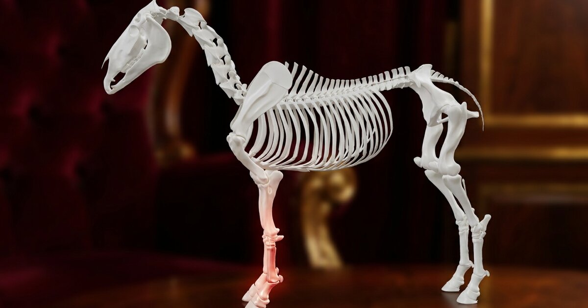
You are hosing off after a normal ride when your hand runs down the back of the cannon bone and stops. Something is puffy back there, and it feels warm. You step back, and the clean straight line of the leg is replaced by a soft outward curve. That curve is where the injury gets its old barn name. A bowed tendon is one of the most common and most heartbreaking soft tissue injuries in horses, partly because it heals so slowly and partly because it loves to come back. Understanding what actually failed inside that leg is the first step to a real recovery. For more details, see our guide on Tendon vs Ligament Injuries in Horses. You may also find our article on Horse Anatomy helpful.
What is a bowed tendon, exactly?
A bowed tendon is damage to the superficial digital flexor tendon, almost always in a front leg. The word "bow" describes the appearance, not a diagnosis. When the tendon tears and the leg swells, the back of the cannon takes on a rounded, convex shape that looks like an archer's bow from the side. Vets call the condition tendinitis or tendinopathy of the SDFT. You may also find our article on Equine Influenza helpful. You may also find our article on EPM in Horses helpful.
The SDFT is the single most commonly injured tendon in the horse. Between 75 and 95 percent of tendon injuries in performance horses involve it, and the forelimbs take the brunt. Thoroughbred racehorses top the statistics, but any horse asked to gallop, jump, or work hard on variable footing can bow a tendon.
Meet the SDFT: the spring in your horse's leg
The superficial digital flexor tendon is essentially a biological spring. It runs from the superficial digital flexor muscle above the knee, down the back of the cannon bone and over the fetlock, then splits and attaches to the pastern. Its job is to flex the lower leg and help support the fetlock as the horse loads the limb.
Here is what makes it so vulnerable. During a gallop, the SDFT stretches by more than 10 percent of its length, storing and releasing energy like a rubber band to help fling the leg forward. That elastic recoil is efficient, but it means the tendon works close to its mechanical limit every single stride. It is often compared to the human Achilles tendon, and anyone who has torn an Achilles knows how that goes. It is built from parallel type I collagen fibers, and once that architecture tears the horse cannot simply rebuild it, which explains most of what is hard about recovery.
SDFT vs DDFT: what is the difference?
Horses have two flexor tendons running down the back of the lower leg, and people mix them up constantly. The SDFT sits closest to the surface, which is why a bow is easy to see and feel. The deeper DDFT lies in front of it, closer to the bone, and does a related but different job. They fail in different ways.
| Feature | SDFT (Superficial) | DDFT (Deep) |
|---|---|---|
| Position | Closest to the skin, at the very back of the cannon | Deeper, between the SDFT and the cannon bone |
| Attaches to | The pastern (short pastern bone) | The coffin bone inside the hoof |
| Main role | Energy-storing spring, supports the fetlock | Flexes the toe, stabilizes the lower joints under load |
| Injury nickname | Bowed tendon | Usually just called a DDFT injury or lesion |
| How often it bows | Very common, the classic bow | Uncommon in the cannon region, more often injured inside the hoof or fetlock |
Each tendon also has a supporting check ligament that shares the load, one reason a proper ultrasound scans the whole leg, not just the puffy spot.
How does a tendon bow in the first place?
Most bows are not a single dramatic accident. They are the visible endpoint of damage building quietly for weeks or months. Every hard gallop or big jump causes a little microdamage to collagen fibers, which the tendon normally repairs between workouts. When the workload outpaces that repair, especially in a fatigued tendon, the accumulated weakness reaches a tipping point and a chunk of fibers tears at once. That is the day you find the heat.
Classic risk factors line up with this picture:
- Fatigue and fast work. Tired muscles stop absorbing shock, dumping more load onto the tendon, so many bows happen in the final stretch of a workout or race.
- Hard, deep, or uneven footing. Ground that grabs or gives unpredictably spikes the strain on the SDFT.
- Ramping up too fast. Tendon adapts far more slowly than muscle, so a program that suits the lungs can overload the tendon.
- Conformation and foot balance. A long toe and low heel, or a back-at-the-knee build, increase leverage on the tendon.
- Cold tendons. A tendon worked hard before it is warmed up is stiffer and less forgiving.
A smaller number of bows are acute, caused by a fall, a strike from another hoof, or overreaching, when a hind foot clips a front leg.
What does a bowed tendon look and feel like?
The three classic signs are heat, swelling, and pain on the back of the cannon. In a fresh injury, gentle finger pressure along the tendon produces a flinch. The swelling creates the bow, that soft outward curve most obvious when you crouch and look at the leg in profile.
Lameness is the tricky part. It ranges from barely noticeable to obvious head-nodding, and here is the catch that surprises a lot of owners: the degree of lameness does not reliably tell you how bad the injury is. A horse can bow significantly and jog off looking nearly sound within days, which is exactly why some get worked right back into a serious tear. In a severe case the fetlock may drop lower than normal under weight, because the damaged SDFT can no longer support it. Any new heat, filling, or bowed shape on the back of a leg deserves a call to your vet and a stop to work.
How vets diagnose it: the ultrasound story
Diagnosis starts with hands. Your vet will palpate the tendon along its length, feeling for heat, thickening, pain, and any change in shape, and compare it against the opposite leg. But the real story is on the ultrasound screen. Ultrasonography, usually with a high-frequency probe (7.5 to 15 MHz), is the gold standard for looking inside a bowed tendon.
On the scan, a classic SDFT tear shows up as a darker (hypoechoic) area within the normally bright, uniform tendon. Many bows produce a central "core lesion," a hole of damage in the middle of the tendon that looks like a dark void surrounded by more normal fibers. The vet also measures the cross-sectional area, since an injured tendon swells, and checks how well the fiber pattern lines up. All of this grades the severity and gives a baseline to measure healing against.
Timing matters. A scan done in the first day or two can underestimate the damage, because inflammation is still evolving and the lesion may enlarge over the following week, so many vets rescan after about a week to map its true size. From there, repeat ultrasounds every two to three months become the roadmap for the whole rehab, telling you when it is genuinely safe to add work rather than guessing from how the horse feels.
The healing timeline: why tendons take so long
Tendon heals in three overlapping phases, and none of them can be rushed. Understanding them makes the long timeline make sense.
Phase 1: Inflammation (roughly the first 1 to 2 weeks)
The body floods the area with inflammatory cells. This is necessary cleanup, but it is a blunt instrument that also damages nearby healthy fibers and can make the lesion look bigger before it looks better. This is the window where icing, cold hosing, anti-inflammatory support, and rest do the most good.
Phase 2: Repair (weeks to a few months)
The tendon fills the gap with a provisional patch built largely from type III collagen, which is quick to lay down but weaker and less elastic than the type I it replaces. In other words, the early repair tissue is scar, not spring.
Phase 3: Remodeling (many months to over a year)
Slowly, the body converts some of that type III collagen to stronger type I and realigns the fibers along the lines of stress. This is where controlled loading matters, because tendon fibers organize themselves in response to gentle, progressive strain. Even so, the repaired tendon typically never fully regains its original strength or elasticity, staying a little stiffer and weaker, which is the core reason bows re-injure.
Rehab: why controlled exercise beats total rest
The biggest mistake with a bowed tendon is doing too much too soon or, surprisingly, doing nothing at all. A tendon left in a stall with zero controlled loading heals into disorganized, weak scar tissue, while one reintroduced to careful, gradually increasing work heals into better-aligned, stronger tissue. Repeat ultrasounds are how you walk that line.
A typical controlled-exercise program, tailored by your vet to the injury, looks roughly like this:
- Weeks 1 to 3: strict stall rest while inflammation settles, then short, controlled hand-walking on flat, firm ground.
- Months 1 to 3: steadily increasing hand-walking, with light trotting usually introduced around three months if the ultrasound looks good.
- Months 4 to 7: longer, more structured work that builds the tendon's tolerance under veterinary monitoring.
- Months 7 to 12: cantering is generally held off until around 7 to 8 months, with a return to full training or competition often between 8 and 12 months.
Plan on a minimum of six months even for a milder bow, and closer to a year for a significant one. Your vet may also discuss regenerative options such as stem cell therapy, platelet-rich plasma, or shockwave, which aim to nudge the tendon toward higher-quality type I collagen repair. Injecting corticosteroids directly into the tendon is contraindicated, because it interferes with repair.
Then there is the number nobody likes. Re-injury is common. Studies of horses treated conventionally report new injuries in roughly 23 to 67 percent within two years, and in racing Thoroughbreds the figure has been reported as high as around 80 percent. There is a hopeful thread: in one study of horses treated with bone-marrow-derived stem cells, the re-injury rate fell to about 27 percent versus roughly 56 percent for conventional management. Patience, honest ultrasound checkpoints, and resisting the urge to bring your horse back early are the things most within your control.
Frequently Asked Questions
Can a bowed tendon heal completely?
The tendon heals and can become sound and functional, but it rarely returns to 100 percent of its original strength and elasticity. It fills in with repair tissue that is partly scar, so the goal of rehab is the strongest, best-organized healing possible. Many horses return to full work with careful management.
How long is a horse out with a bowed tendon?
Expect a minimum of six months and often 8 to 12 months of controlled rehabilitation for a significant bow. The timeline depends on the size of the lesion and how the tendon looks on repeat ultrasounds. Rushing the return is the fastest way to end up back at the start.
Should I hand-walk a horse with a bowed tendon or rest it completely?
Both, in the right order and only on your vet's schedule. The first week or so is usually strict rest to calm inflammation, followed by carefully controlled hand-walking. Progressive loading helps the fibers heal in a stronger, more organized pattern than total stall rest alone.
Is a bowed tendon the same as a strained deep flexor tendon?
No. A bow specifically refers to the superficial digital flexor tendon (SDFT), the one closest to the skin. The deep digital flexor tendon (DDFT) is deeper and attaches to the coffin bone inside the hoof, and it is injured less often in the cannon region. Only ultrasound can confirm which structure is involved.
Why do bowed tendons come back so often?
Because the healed tendon is a little weaker and stiffer than the original, so it is more likely to fail again under hard work, especially if the horse returns too soon. Re-injury rates of 23 to 67 percent within two years have been reported with conventional treatment. Slow, monitored rehab is the best defense. For more details, see our guide on Thrush in Horses.
Sources
- Merck Veterinary Manual. Tendinitis in Horses. Musculoskeletal System: Tendon and Ligament Disorders in Horses. merckvetmanual.com.
- Baxter, G.M. (Ed.) (2020). Adams and Stashak's Lameness in Horses, 7th Edition. Wiley-Blackwell.
- Lesté-Lasserre, C. Understanding Equine SDFT Injuries. The Horse. thehorse.com.
- Godwin, E.E., Young, N.J., Dudhia, J., Beamish, I.C., & Smith, R.K.W. (2012). Implantation of bone marrow-derived mesenchymal stem cells demonstrates improved outcome in horses with overstrain injury of the superficial digital flexor tendon. Equine Veterinary Journal, 44(1), 25-32.
- Avonvale Equine Practice. A Guide to the Superficial Digital Flexor Tendon. avonvaleequine.co.uk.
- Mad Barn. Bowed Tendons in Horses: Tendinitis Clinical Signs and Treatment. madbarn.com.