Horse Anatomy: A Complete Reference Guide |
Most horse anatomy guides are a list of parts.
This one is organized around a different question: what does each system actually do, and what does that mean for the horse standing in your barn? Because the reason the digestive tract is thirty feet longer than it needs to be, and the reason the front legs are not attached to the skeleton at all, are the reasons horses colic and go lame.
Every structure named here is clickable in the Inside The Equine 3D explorer, where you can rotate the horse, strip away layers, and look at the thing itself instead of a drawing of it.
Quick Answer
The horse's body is organized into several major systems. The skeletal system contains roughly 205 bones. The muscular system is the largest tissue mass in the body and includes the thoracic sling, which suspends the rib cage between the forelimbs, because horses have no collarbone. Tendons and ligaments do the work of muscle below the knee and hock, where there is no muscle at all. The digestive system runs roughly 100 feet and is a hindgut fermenter built for continuous grazing. The hoof contains the coffin bone, suspended inside the hoof capsule by the laminae. Understanding these systems is what makes equine health conditions comprehensible rather than mysterious.
The Skeletal System
The adult horse has approximately 205 bones.
Say "approximately" and mean it. That number is the standard figure and it is the one you will see everywhere, but it genuinely varies. Some Arabians have a shorter thoracic spine and carry 17 ribs rather than the usual 18, and bones fuse over the course of a life, which changes the count. Anyone who tells you a horse has exactly 205 bones is repeating a number rather than describing an animal.
The skeleton divides into two parts:
- The axial skeleton: skull, vertebral column, sternum, ribs
- The appendicular skeleton: the limbs
The spine
Seven cervical (neck) vertebrae, the same number as a human and a giraffe. Around 18 thoracic vertebrae, though 17 to 19 occurs. Roughly six lumbar. Five sacral vertebrae, fused. Then 15 to 21 caudal vertebrae in the tail.
The withers are the spinous processes of the thoracic vertebrae. That bony ridge you rest your hand on is the top of the spine itself, poking upward. It is worth knowing because it explains why saddle fit is an anatomical problem rather than an upholstery problem, and why kissing spines happens where it happens.
The ribs
Around 18 pairs. Eight of them are "true" ribs, meaning they connect all the way from the spine to the sternum. The rest are "floating," attached to the spine but not reaching the sternum directly.
The forelimb is not attached to the skeleton
This is the single most surprising fact in equine anatomy, and it explains an enormous amount.
A horse has no collarbone. In a human, the clavicle physically bolts the arm to the trunk. The horse has nothing there. Instead, the forelimb is suspended from the body entirely by muscle, tendon and fascia, in a structure called the thoracic sling. The rib cage hangs in a muscular hammock between the two shoulder blades.
The consequences are everywhere. It gives the horse enormous shoulder mobility and lets him fold his front legs tightly over a jump. It also means that if the thoracic sling musculature is weak, the rib cage sinks between the shoulder blades, and the whole posture of the horse changes. Most of what a rider is trying to develop when they talk about "lifting through the shoulder" is this.
The hind limb, by contrast, is bolted on, through the pelvis to the spine. The hind end carries roughly 40 percent of the weight but produces most of the forward movement. The front end carries about 60 percent and absorbs most of the concussion, which is precisely why front limb lameness is so much more common.
Each limb contains roughly 20 bones. Sources differ slightly on the exact count for the hind limb, which is a good reminder that anatomy is a description of biology, not arithmetic.
The Muscular System
Muscle is the largest tissue mass in the horse's body. There are three types: skeletal muscle, which moves the horse and which you can train; smooth muscle, which lines the gut and blood vessels and works involuntarily; and cardiac muscle, which exists only in the heart.
The groups that matter most to a rider:
- The hindquarter. Croup, thigh, gaskin. This is the engine. Propulsion comes from here.
- The topline. Longissimus dorsi and the muscles along the spine. This is what carries a rider.
- The thoracic sling. Abdominal muscles, chest muscles, and the "wither lifters." This is what holds the rib cage up between the front legs and it is the foundation of sound ridden work.
- The neck and shoulder. Brachiocephalicus, splenius, trapezius.
One fact reframes everything about conditioning: muscle adapts to training throughout the horse's life. Tendon largely does not. That asymmetry is the whole logic behind strengthening a horse's legs and tendons, and it means the muscle you build above the leg is the thing protecting the leg you cannot build.
Tendons and Ligaments
The distinction is simple and worth getting right, because owners use these words interchangeably and vets do not.
Tendons connect muscle to bone. They transmit force.
Ligaments connect bone to bone. They stabilize joints.
Both are made of collagen. Both have a poor blood supply, which is why both heal slowly, and why an injury to either is measured in months rather than weeks.
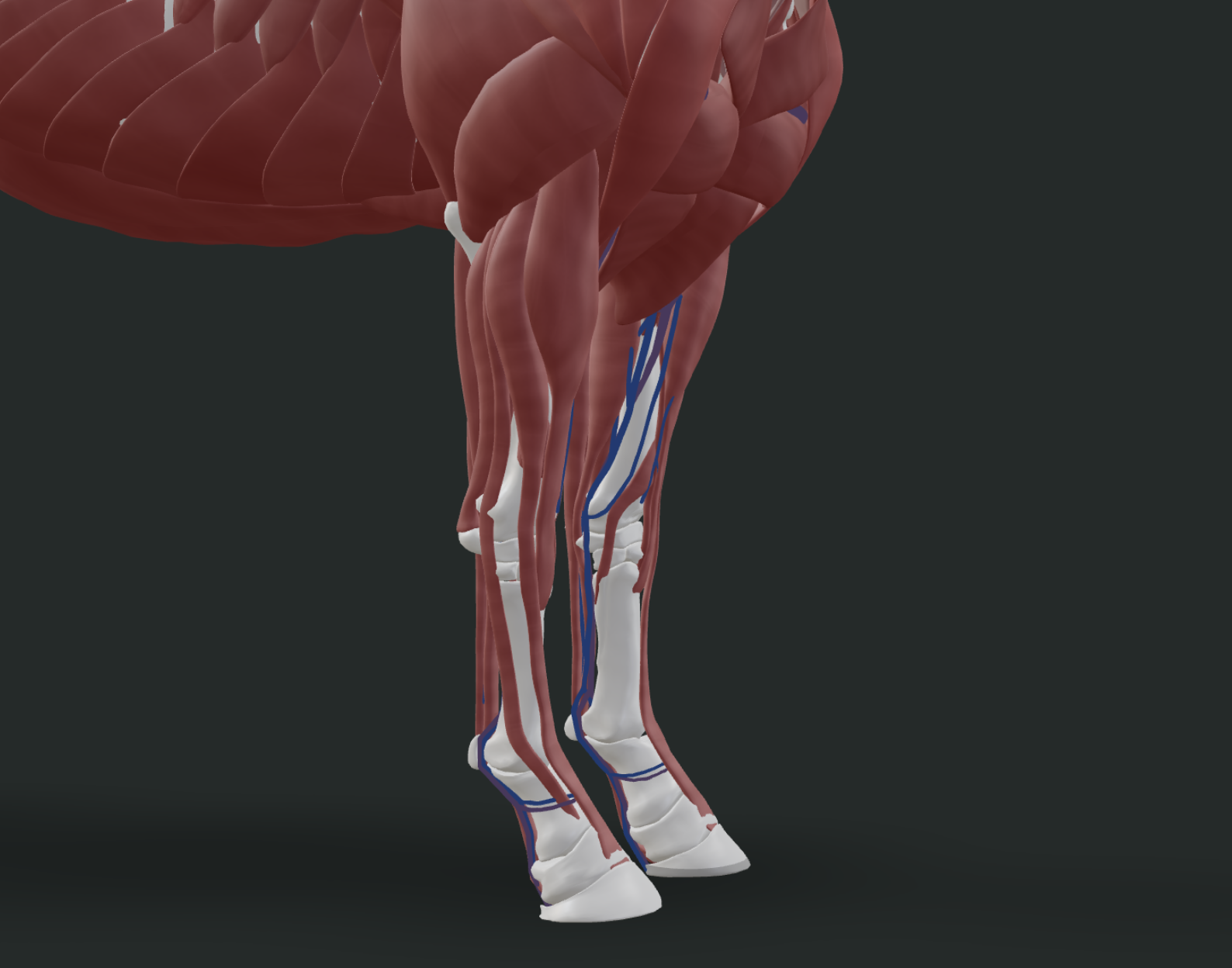
Below the knee, there is no muscle
None. From the carpus down, the horse's leg is bone, tendon, ligament, joint capsule and skin. This is the fact that everything about lower limb health hangs on.
The main players:
- Superficial digital flexor tendon (SDFT). Runs down the back of the cannon bone. An elastic, energy-storing tendon. The classic site of a bowed tendon.
- Deep digital flexor tendon (DDFT). Runs beneath the SDFT, continues around the navicular bone, attaches to the coffin bone.
- Suspensory ligament. Runs down the back of the cannon beneath the flexor tendons, splits into two branches attaching to the sesamoid bones. It stops the fetlock from dropping to the ground under load.
- Check ligaments. Limit how far the flexor tendons can stretch.
- The nuchal and supraspinous ligament system. Runs from the poll all the way back along the spine to the sacrum, a single connected elastic system linking the horse's head carriage to his back.
Because there is no muscle down there, there is also no muscle pump to push fluid back up the leg. Fluid return depends on movement and on the compression of the frog with every stride. That is exactly why horses stock up and get swollen legs when they stand still.
The full picture of what lives in the lower limb, and what goes wrong with each structure, is in the guide to horse leg injuries and rehabilitation.
The Hoof and Lower Limb
The horse stands on the equivalent of a single fingertip. The hoof is a modified third digit, and the hoof wall is homologous to your fingernail.
Inside the hoof capsule:
- The coffin bone (P3, third phalanx). The bone inside the hoof. It is suspended, not resting, inside the capsule.
- The laminae. Interlocking, velcro-like soft tissue folds that suspend the coffin bone from the inner hoof wall. When the laminae fail, the coffin bone rotates or sinks. That is laminitis. The entire disease is one sentence of anatomy.
- The navicular bone. A small sesamoid bone sitting behind the coffin joint, with the DDFT running over it like a rope over a pulley. Pain in this region is navicular syndrome.
- The digital cushion and the frog. Shock absorption, and the pump that drives circulation back up the leg on every stride.
- The proximal sesamoid bones. Two small bones at the back of the fetlock, where the suspensory ligament branches attach.
The hoof anatomy guide goes deeper, and the hoof growth rate post covers what that structure does over time.
The Digestive System
The horse's digestive tract is roughly 100 feet long, and most of it is intestine.
This is the anatomy of an animal evolved to walk across a semi-arid plain eating small amounts of poor-quality grass, all day, every day. Nearly every digestive problem in a domestic horse traces back to the gap between that design and how we actually keep horses.
Running the length of it:
- The mouth and teeth. Grinding surfaces that wear continuously and erupt continuously to compensate.
- The esophagus. About four to five feet. This is where choke happens.
- The stomach. Remarkably small: around 19 litres (roughly 5 gallons) in an average horse, and it functions best when only about a third full. It empties when it is around two thirds full whether digestion has finished or not, which is the anatomical argument for small frequent meals and continuous forage.
- The small intestine. 50 to 70 feet. Where most enzymatic digestion and absorption happens.
- The cecum and large colon. The fermentation vat. This is where microbes break down fiber. It is the reason a horse can live on grass and it is the reason a horse colics.
Why horses cannot vomit
Two anatomical features. The cardiac sphincter, the muscular ring between the esophagus and the stomach, is exceptionally well developed in horses. And the esophagus meets the stomach at an oblique angle, which effectively creates a one-way valve.
The result is that a horse physically cannot relieve gastric pressure by vomiting. That single structural fact is why colic is a life-threatening emergency in a horse and an unpleasant afternoon in a dog.
The Circulatory and Respiratory Systems
The equine athlete is an extraordinary aerobic machine, and the plumbing explains it.
Circulation. The heart is cardiac muscle, working involuntarily and continuously. The spleen deserves special mention: it stores red blood cells and releases them into circulation during exertion, effectively giving the horse a built-in blood transfusion the moment he starts to gallop.
Respiration. Horses are obligate nasal breathers. They cannot breathe through their mouths. Air goes in through the nostrils, through the nasal passages, past the guttural pouches, through the larynx and trachea, and into the lungs.
At the canter and gallop, breathing is mechanically locked to stride. One breath, one stride. The viscera slide forward and back inside the abdomen like a piston, driving the diaphragm. A horse cannot breathe faster without moving faster, which is a constraint no other athlete has to live with.
The Head, Teeth and Senses
The skull is a complex of many bones, most of them firmly fused. The lower jaw, the mandible, is the significant moving exception.
Teeth. Numbers vary by sex and by individual. Stallions and geldings typically carry 40 to 44 teeth, mares typically 36 to 40, the difference being that mares often do not develop canine teeth. Horse teeth erupt continuously through life as the grinding surface wears down, which is why dental care is not optional and why a horse's age can be roughly estimated from its teeth.
The eyes. Set on the sides of the head, giving nearly panoramic vision with two blind spots: directly in front of the nose, and directly behind. The horse is a prey animal and the anatomy of its head is the anatomy of something built to see a predator coming from any direction.
The guttural pouches. Two large air-filled sacs connected to the eustachian tubes, unique to equids, sitting where major arteries and nerves pass through. Nobody is entirely certain what they are for.
How to Actually Learn This
Reading a list of structures is the least effective way to learn anatomy, and every horse person who has tried to memorize a poster already knows it.
Anatomy is spatial. The suspensory ligament makes no sense as a paragraph and immediate sense the moment you see it running under the flexor tendons and splitting around the back of the fetlock. The reason laminitis is terrifying only lands when you see how the coffin bone actually hangs inside the hoof.
That is the whole reason Inside The Equine exists. Rotate the horse. Turn the muscle layer off. Click the structure and see what it is called and what it does. Then go out to the barn and put your hand on it.
The courses work through each system in order, and the conditions library connects each structure to what goes wrong with it.
Frequently Asked Questions
How many bones does a horse have?
An adult horse has approximately 205 bones. The number varies between individuals and breeds. Some Arabians, for example, have a shorter thoracic spine and 17 ribs rather than the more typical 18. Bones also fuse over the course of a horse's life, which changes the count. 205 is the standard reference figure, not a fixed rule.
Why can't horses vomit?
Two anatomical reasons. The cardiac sphincter between the esophagus and the stomach is exceptionally well developed in horses, and the esophagus enters the stomach at an oblique angle, which together act as a one-way valve. Because a horse cannot relieve gastric pressure by vomiting, colic is a genuine emergency in horses rather than a passing illness.
Does a horse have a collarbone?
No. The horse has no clavicle, so the forelimb is not attached to the skeleton by bone at all. It is suspended from the trunk entirely by muscle, tendon and fascia, in a structure called the thoracic sling. The rib cage effectively hangs in a muscular hammock between the two shoulder blades.
Is there muscle in a horse's lower leg?
No. Below the knee and the hock, the horse's leg contains bone, tendon, ligament, joint capsule and skin, and no muscle. This keeps the limb light and fast, but it means the lower leg has no muscle pump to return fluid upward, and no muscular shock absorption. It is the single most important fact in lower limb health.
How long is a horse's digestive tract?
Roughly 100 feet, and most of that is intestine. The small intestine alone runs 50 to 70 feet. The horse evolved as a continuous grazer of low-quality forage and is a hindgut fermenter, digesting fiber through microbial fermentation in the cecum and large colon.
What is the coffin bone?
The coffin bone, also called P3 or the third phalanx, is the bone inside the hoof capsule. It does not rest on the bottom of the hoof. It is suspended from the inner hoof wall by interlocking soft tissue folds called the laminae. When those laminae fail, the coffin bone rotates or sinks, which is what laminitis is.
Want to go deeper into horse anatomy?
Our Anatomy course breaks down every body system with detailed visuals and quizzes.
Start the Anatomy CoursePro members only. Try Pro free for 7 days.