Navicular Disease in Horses: A Deep Dive Into Causes, Signs, and Management
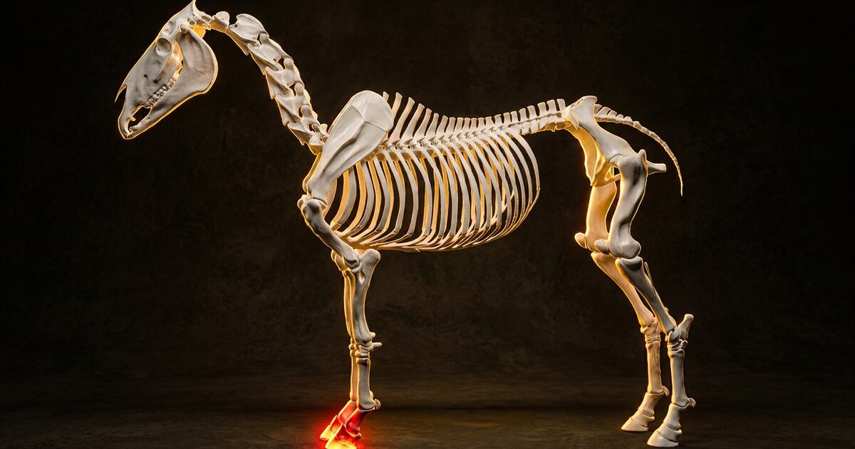
Few words land harder in a barn aisle than "navicular." For decades it was treated as a death sentence for a riding career. The reality today is more hopeful. We now know navicular is not one problem but a whole cluster of injuries in a tiny, hardworking region at the back of the foot. The catch is that the earlier you catch it, and the more precisely you understand what is going wrong, the better your odds. You may also find our article on Horse Anatomy helpful. You may also find our article on Equine Influenza helpful.
What is navicular disease in horses?
Navicular disease is a degenerative condition affecting the navicular apparatus, the collection of structures at the back and bottom of the coffin joint inside the hoof. Vets increasingly call it navicular syndrome or podotrochlosis, and that shift in language matters. "Disease" implied one specific illness of one bone. "Syndrome" and "podotrochlosis" acknowledge what imaging has taught us: the pain can originate from bone, soft tissue, a bursa, or several at once, and no two horses are quite identical. For more details, see our guide on Cushing's Disease in Horses (PPID). You may also find our article on EPM in Horses helpful.
It is genuinely common. Navicular-related pain is estimated to account for up to one-third of all chronic forelimb lamenesses in horses. It shows up most in the front feet, which carry roughly 60 percent of the horse's body weight and absorb enormous force with every stride.
The anatomy: navicular bone, bursa, and deep digital flexor tendon
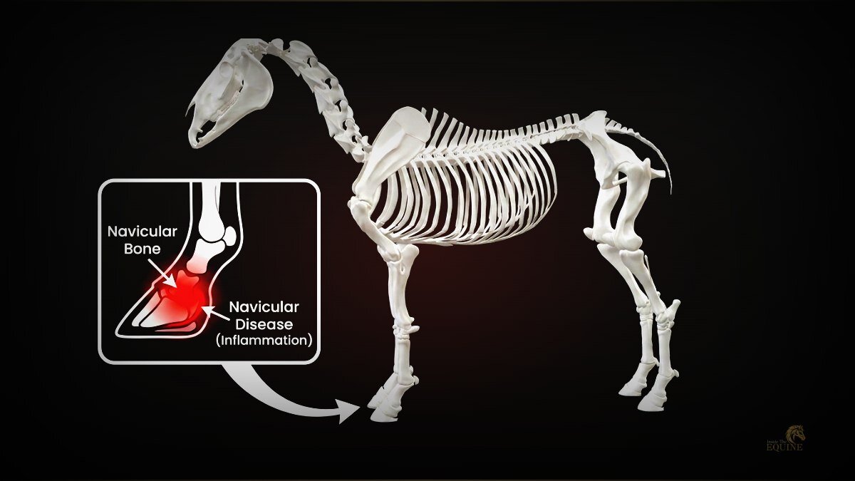
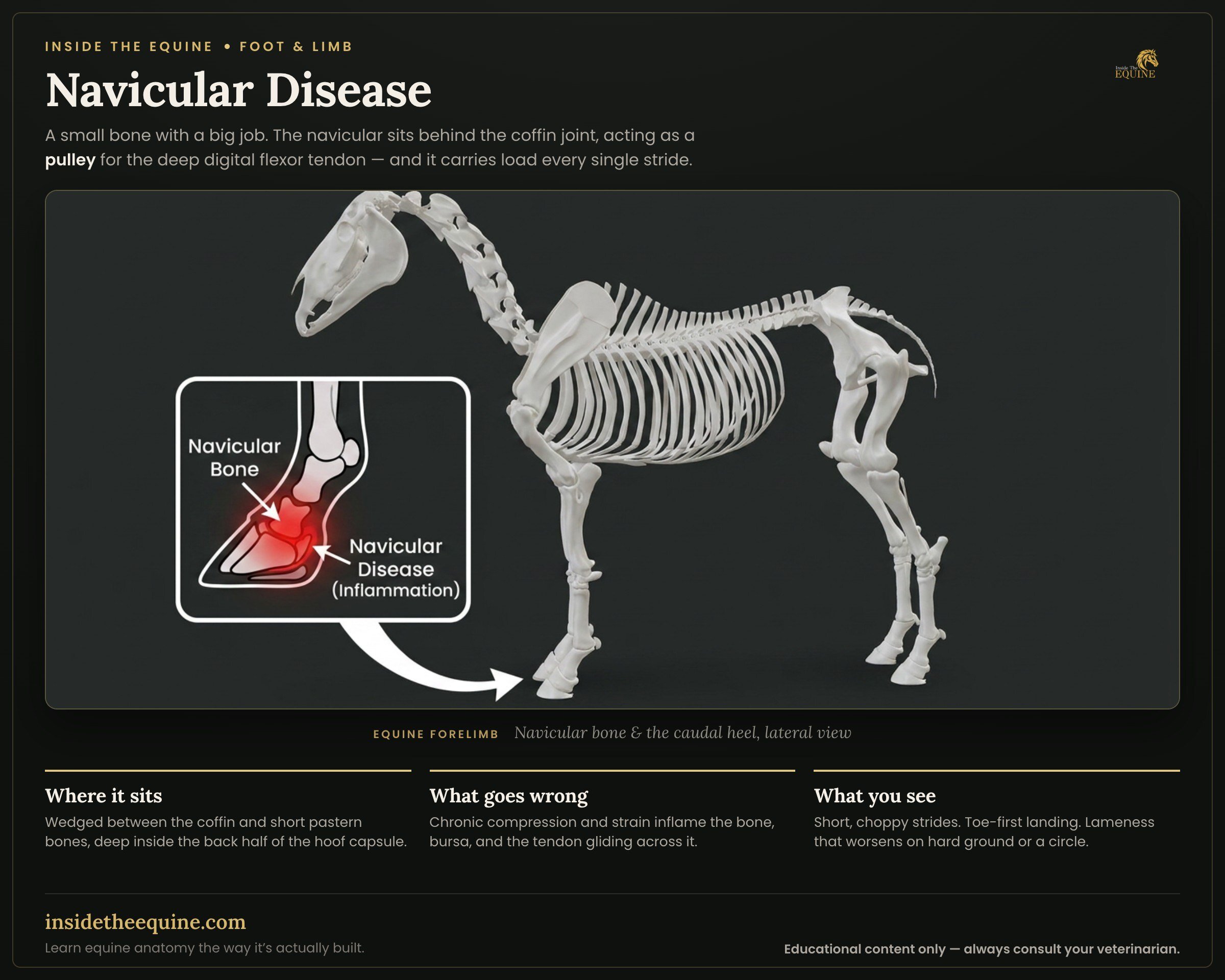
Picture the neighborhood it lives in. The navicular bone is a small, boat-shaped bone (its name comes from the Latin for "little ship") that sits behind the coffin joint, wedged between the coffin bone and the short pastern bone. It acts as a fulcrum, or pulley point.
Draped over the back of that pulley is the deep digital flexor tendon, or DDFT, a powerful tendon that runs down the back of the leg, wraps under the navicular bone, and attaches to the underside of the coffin bone. Every stride, the DDFT slides across the navicular bone under load. To stop those two surfaces from grinding each other raw, a small fluid-filled cushion called the navicular bursa sits between them. Holding the assembly in place are two key ligaments: the collateral sesamoidean ligaments up top and the distal impar ligament below.
Here is the key point: damage to any one of those structures can produce the same lameness. A horse can have a pristine navicular bone but a torn DDFT, or a healthy tendon but an inflamed bursa and eroded bone. Research consistently finds that many horses with navicular bone changes also have DDFT lesions, which tells us these injuries feed off each other and compound over time.
What causes navicular disease?
There is no single cause, which is why it took so long to understand. Think of several forces stacking up over time, mostly some combination of conformation, biomechanics, and genetics.
Conformation and hoof shape
The classic at-risk foot is upright and narrow with contracted heels and a small frog, or the opposite extreme of a long toe with an underrun, collapsed heel. Both shapes shift force onto the back of the foot in ways nature did not intend. A large body on disproportionately small feet also loads a small footprint with a lot of mass, concentrating stress on the navicular region.
Biomechanics and the long-toe lever
This is the mechanism farriers care about most because it is the one they can change. When the toe grows long and the heel runs low, the breakover point moves forward and the coffin joint's center of rotation shifts toward the back of the hoof. That creates a longer lever arm, increasing the tension the DDFT exerts as it curves around the navicular bone, which means more compression on the bone and bursa every stride. Add hard footing, tight turns, sliding stops, and jumping, and you multiply the strain.
Genetics and vascular factors
Heredity is a real risk factor, which is why certain breeds are overrepresented: American Quarter Horses (often bred for small, neat feet on big muscular bodies), Warmbloods, and Thoroughbreds. Some researchers also point to altered blood flow to the navicular bone and chronic inflammation resembling osteoarthritis. In young horses, structural bone defects can exist without lameness, only turning painful years later as the surrounding soft tissue gives way.
What are the signs of navicular disease?
The hallmark sign is a subtle, chronic, low-grade lameness in both front feet that owners describe as the horse not moving right. Because it is usually bilateral, you may not see obvious head-bobbing on a straight line. Instead the horse looks stiff or "pottery," and one foot is often worse than the other.
Watch for these patterns:
- A short, choppy, "stabby" stride that gets worse on hard ground and on tight turns.
- Toe-first landing. A sound horse lands heel-first or flat, but a horse with heel pain drops the toe first to spare the sore back of the foot.
- Stumbling, shuffling, or reluctance to move forward freely.
- Intermittent lameness that warms out early, then returns, or comes and goes week to week.
- Pointing a front foot at rest, or shifting weight between the front feet at the crossties.
- Sensitivity when hoof testers are applied across the frog and heel region.
Because these signs are vague and develop slowly, navicular is easy to dismiss as the horse being lazy or "just needing more fitness." That misread costs time. Persistent, low-grade front-end lameness deserves a real workup.
How do vets diagnose navicular disease?
Diagnosis means localizing the pain first, then imaging to see what is causing it. No single test does the whole job.
The first step is usually a lameness exam with flexion tests and hoof testers, followed by diagnostic nerve blocks. A palmar digital nerve block numbs the back third of the foot. If the horse goes noticeably sounder afterward, often around a 90 percent improvement in a positive case, that points strongly to pain in the heel region. This is localization, not yet a diagnosis: the block cannot tell bone from tendon from bursa.
To see the structures themselves, vets turn to imaging. Each tool shows something different:
| Imaging tool | What it shows best | Limitations |
|---|---|---|
| Radiographs (X-ray) | Bone changes: enlarged vascular channels, cyst-like lesions, sclerosis, bone spurs, flexor cortex erosion | Cannot see soft tissue. Slight angle changes alter the picture, so reproducible views matter |
| MRI | The gold standard. Shows bone and every soft tissue at once: DDFT tears, bursa inflammation, ligament damage | Expensive and may require general anesthesia or a standing MRI facility |
| Ultrasound | Can catch DDFT lesions extending up into the pastern | Limited inside the hoof capsule; low overall diagnostic yield for the navicular region |
Radiographs remain the practical workhorse because they are affordable and let a vet track bone changes over time. But MRI is what genuinely changed the game. By showing the DDFT, bursa, and ligaments along with the bone, it lets a vet say which structure is actually driving the lameness. This is why "navicular" today is really a starting point for a more specific diagnosis rather than a final answer.
How is navicular disease managed?
There is no cure, but there is a lot of effective management. The guiding principle is clear: start with the mechanics of the foot, and treat medication as a complement rather than the main event. You cannot inject your way out of a badly balanced foot.
Corrective farriery first
The foundation is corrective shoeing that eases the load on the navicular apparatus. The goals are to shorten the toe, support and often modestly elevate the heel (commonly a couple of degrees, adjusted to the individual foot), and ease breakover with a rolled or rockered toe. Reducing that long-toe lever directly lowers the tension the DDFT places on the navicular bone. Many vets and farriers use before-and-after radiographs to confirm the trim and shoe are genuinely improving the foot's geometry.
Medication and injections
Layered on top of good farriery, medical options include:
- Anti-inflammatory drugs (NSAIDs) such as phenylbutazone or firocoxib to control pain and inflammation.
- Intra-synovial corticosteroid injections, sometimes with hyaluronic acid, into the coffin joint or the navicular bursa itself.
- Systemic bisphosphonates (tiludronate or clodronate), which target bone remodeling and are labeled for navicular-associated bone pain.
- Regenerative or biologic therapies (platelet-rich plasma, autologous conditioned serum, or stem cells) where soft tissue damage is involved, plus extracorporeal shock wave therapy as an adjunct.
Dosing and drug choice are strictly your veterinarian's call, tailored to what the imaging shows and how the horse responds. Two horses labeled "navicular" can need completely different plans depending on whether the problem is mainly bone or tendon.
Surgery and last resorts
When medical and farriery management stop working, surgical options exist. Navicular bursoscopy is a minimally invasive procedure to debride DDFT tears and bone erosions within the bursa. A palmar digital neurectomy, which cuts the sensory nerves to the back of the foot, is reserved for cases that have exhausted other options and have minimal tendon damage. It relieves pain but fixes nothing, the nerves usually regrow within one to two years, and the loss of sensation means the horse can no longer feel a hoof abscess. It is a serious decision to make only with your vet.
Can a horse with navicular disease keep working?
Often yes, and this is where the modern outlook diverges most from the old reputation. The prognosis is generally described as guarded to fair, and navicular is progressive, requiring lifelong management. Some horses eventually stop responding to treatment. But many others, caught early and managed well, stay comfortable and in work for years, sometimes dropping to a less concussive discipline rather than retiring outright. For more details, see our guide on Thrush in Horses.
The single biggest lever you control is time. Front-end lameness that lingers past a couple of weeks, a horse landing toe-first, a stride that shortens on hard ground: these are worth a call to your vet sooner rather than later. Navicular rewards early, specific diagnosis and punishes wishful thinking. A horse managed thoughtfully from the first warning signs has a very different future than one whose owner waited a year hoping it would sort itself out.
Frequently Asked Questions
Is navicular disease curable?
No, navicular disease cannot be cured, because it involves degeneration of bone and soft tissue that does not fully reverse. It can, however, be managed. With corrective farriery, medication, and workload changes, many horses stay comfortable and in work for years. The condition is progressive, so ongoing management is the realistic goal.
What are the first signs of navicular in a horse?
Early signs are usually subtle: a short, choppy stride that worsens on hard ground and tight turns, intermittent lameness in both front feet, and a tendency to land toe-first instead of heel-first. Because these signs are vague and often bilateral, they are easy to mistake for laziness or lack of fitness.
Which breeds are most prone to navicular disease?
American Quarter Horses, Thoroughbreds, and Warmbloods are the most commonly affected breeds. Quarter Horses in particular are often bred for small, neat feet on large muscular bodies, which concentrates load on a small footprint. Genetics is considered a real risk factor, so conformation traits that run in these breeds contribute to the pattern.
Why is MRI important for diagnosing navicular?
MRI is the gold standard because it shows both the bone and all the soft tissue structures at once, including the deep digital flexor tendon, the navicular bursa, and the ligaments. X-rays only reveal bone changes, so a horse can look normal on radiographs but have a torn tendon that only MRI detects. Knowing exactly which structure is damaged lets your vet build a far more targeted treatment plan.
Can good farriery prevent or slow navicular disease?
Balanced trimming and shoeing are central to both prevention and management, though they cannot override genetics. Keeping the toe from getting long, supporting the heel, and easing breakover reduce the leverage the deep digital flexor tendon places on the navicular bone. Regular farrier care on a consistent schedule is one of the most practical things an owner can do.
Is navicular disease the same as heel pain?
Not exactly. Navicular disease is one specific cause of caudal (back-of-foot) heel pain, but heel pain can also come from bruising, soft tissue injury, or coffin joint problems. That is why vets localize the pain with a nerve block first, then use imaging to determine whether the navicular apparatus is truly the culprit.
Sources
- Merck Veterinary Manual. Navicular Syndrome in Horses. Musculoskeletal System, Disorders of the Foot in Horses. Merck & Co.
- Utah State University Extension. Equine Navicular Syndrome. USU Extension Equine Program, 2023.
- Lesté-Lasserre, C. Diagnosing and Managing Equine Navicular Syndrome (featuring Sammy Pittman, DVM). The Horse.
- The Horse. A Fresh Look at the Causes of Navicular Disease. TheHorse.com.
- Baxter, G.M. (Ed.). Adams and Stashak's Lameness in Horses (7th Ed.). Wiley-Blackwell.