How to Study Horse Anatomy on Your Phone
You are standing in a vet appointment, nodding along while the doctor points to an X-ray and says something about the navicular bone and the deep digital flexor tendon. You know roughly where the hoof is. Beyond that, you are faking it.
That moment, the one where you smile and pretend you followed every word, is exactly why studying horse anatomy matters. Not to become a vet. To become a better advocate for your horse.
The good news? Your phone is a better anatomy classroom than most college textbooks ever were. Here is how to actually use it.
Why Bother Learning Anatomy at All?
Skip this section if you already know the answer. For everyone else: anatomy knowledge changes how you communicate with professionals and how quickly you catch problems.
When your farrier mentions the hoof-pastern axis, you want to know whether that is about angles or tendons (it is both). When your horse comes in from the pasture short on the left front and you can feel heat in the hock, you need to know whether that heat matters or whether hocks just run warm sometimes. Spoiler: it matters.
Horse owners who understand basic anatomy catch lameness earlier, describe symptoms more precisely, and make faster decisions in emergencies. A study published in the Journal of Veterinary Medical Education found that visual and interactive learning tools significantly improved retention of anatomical concepts compared to textbook-only study. Your phone gives you access to those tools everywhere you go.
Start With the Skeleton, Not the Muscles
This is the mistake almost everyone makes. They jump straight into muscles and tendons because those feel more "practical." But muscles attach to bones, tendons cross joints, and ligaments hold everything together. Without the skeletal map in your head, the soft tissue is just a pile of Latin words.
Start by learning the major bones of the horse's skeletal system. There are 205 of them, give or take depending on the source, but you do not need all 205. Focus on the landmarks that vets and farriers actually reference:
- The scapula and its relationship to shoulder angle
- The radius, carpus (knee), and cannon bone down the front leg
- The hock and its complex stack of tarsal bones
- The coffin bone and navicular bone inside the hoof
- The pelvis, sacroiliac joint, and femur in the hindquarters
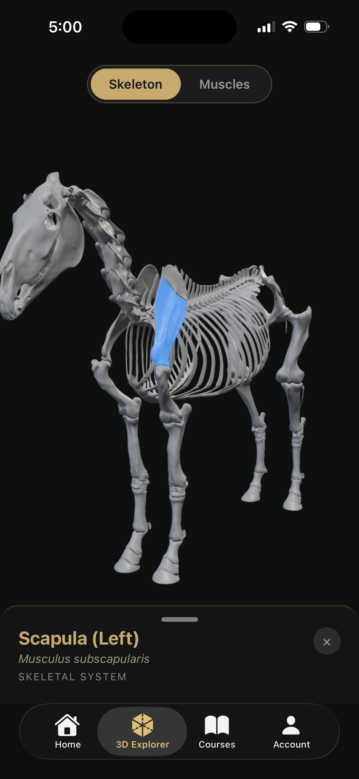
The Inside The Equine app is built specifically for this kind of exploration. You can rotate a 3D horse skeleton, tap any bone, and read what it does, all without digging through a 400-page textbook. That kind of spatial understanding is hard to get from flat diagrams.
Use 3D Models to Build Spatial Memory
Here is a thing about anatomy that textbooks get wrong: the horse is not flat. The stifle sits at an angle. The scapula slopes backward. The navicular bone hides behind the coffin bone where you cannot see it from the outside.
Two-dimensional diagrams force your brain to reconstruct three dimensions from a flat image. That is hard cognitive work, and it is why so many people study anatomy diagrams, pass the quiz, and forget everything a week later.
3D models fix this. When you can spin a hock joint around and look at it from the medial side, the lateral side, and straight on, the spatial relationships click in a way that sticks. Research on medical education supports this: a 2020 meta-analysis in Anatomical Sciences Education found that 3D visualization tools consistently outperformed traditional methods for spatial anatomy learning.
The Inside The Equine app gives you this on your phone. Pinch to zoom into the digital flexor tendons. Rotate the suspensory ligament to see where it originates and where it branches. This is the kind of hands-on exploration that builds real understanding.
Connect Anatomy to Real Problems
Studying anatomy in isolation is boring. Studying it because your horse just got diagnosed with navicular syndrome is riveting. Use that energy.
The most effective way to learn anatomy on your phone is to pair every structure with a clinical condition:
- Learning the laminae? Read about laminitis at the same time. Suddenly the anatomy has stakes.
- Studying the stifle joint? Look up locked stifle and stifle injuries. Now those ligament names have meaning.
- Exploring the nuchal ligament? Connect it to kissing spine and how head carriage affects the back.
- Reading about the sacroiliac joint? Understand why "hunter's bump" shows up and what it means for performance.
Inside The Equine's website pairs anatomy pages with related conditions, so you can bounce between structure and pathology without losing your place. This back-and-forth is how veterinary students learn, and it works just as well on your phone during your lunch break.
Build a Daily Study Habit (Even 5 Minutes Works)
You do not need hour-long study sessions. Anatomy retention depends more on frequency than duration. Five minutes a day, every day, beats a two-hour cram session once a month.
Here is a simple routine that works:
Morning (2 minutes): Open the Inside The Equine app and explore one structure you have never looked at before. Read the description. Rotate it. Done.
At the barn (1 minute): Palpate that structure on your actual horse. Can you feel the point of the hock? Can you find the digital pulse? The transition from screen to horse is where learning becomes knowledge.
Evening (2 minutes): Read one blog post or condition page related to what you studied. If you learned about the coffin bone this morning, read about navicular syndrome tonight.
That is five minutes. Over a month, you will have studied 30 different structures and their clinical relevance. Over six months, you will know more functional horse anatomy than most amateur riders learn in a lifetime.
Use Your Phone at the Barn
This is where digital study tools earn their keep. A textbook stays on your shelf. Your phone goes everywhere.
Standing next to your horse with an anatomy app open is the single best way to learn. You can look at the 3D model of the equine eye, then look at your horse's actual eye. You can study the respiratory system, then listen to your horse breathe. You can read about gut sounds, then press your ear to your horse's flank.
This is not abstract studying. It is applied learning, and it is how professionals develop clinical intuition.
A few practical tips for phone-at-the-barn studying:
- Use a waterproof case. Barns are dusty, muddy, and occasionally involve flying water buckets.
- Download content for offline access. Cell service in rural barns is often terrible.
- Bookmark structures you want to palpate. Make a list before you go out, then check them off on your horse.
- Take photos of your horse's legs, hooves, and body. Compare them to the anatomical models. Notice where tendons run, where joints flex, where bones create landmarks you can feel through the skin.
Study With Purpose: Match Your Learning to Your Discipline
A dressage rider and a barrel racer need different anatomical knowledge, even though the horse is the same animal.
If you ride dressage or flat work: Focus on the thoracic sling, the nuchal ligament, shoulder angle, and how collection changes spinal mechanics. Understanding the stay apparatus helps you grasp why some horses struggle to engage their hindquarters.
If you jump: Study the suspensory ligament, tendon vs. ligament anatomy, hock structure, and how the cardiovascular system supports intense effort. Understanding eye position during jumping will change how you approach fences.
If you are a trail rider or general owner: Focus on vital signs, hoof anatomy, the digestive system, and signs of pain. These are the structures and systems most likely to matter in day-to-day horse care.
If you are a breeder: Reproductive anatomy, foal navel care, and the integumentary system for wound assessment are your priorities.
Free and Paid Resources Worth Your Time
Your phone gives you access to more equine anatomy resources than existed in entire university libraries 20 years ago. Here are the ones actually worth using:
Inside The Equine (app and website): Purpose-built for horse people, not adapted from human medical apps. The 3D models are specific to equine anatomy, and the content connects structures to real conditions horse owners face. Available on iOS.
University extension resources: Many land-grant universities publish free equine anatomy guides. Colorado State, UC Davis, and Cornell all have solid online materials.
Veterinary podcasts: Shows like The Horse podcast and Equine Science Update regularly cover anatomy-related topics. Listen during your commute and follow along with a 3D model later.
Social media (with caution): Instagram and TikTok have equine anatomy content ranging from excellent to dangerously wrong. Follow veterinarians and university accounts. Be skeptical of anyone selling miracle supplements alongside their anatomy lessons.
Frequently Asked Questions
Do I need a science background to study horse anatomy?
Not at all. Modern apps and 3D tools are designed for visual learners without any prerequisite knowledge. Start with the skeleton, learn the major landmarks, and build from there. If you can learn your horse's feeding schedule, you can learn where the coffin bone sits.
How long does it take to learn basic horse anatomy?
With consistent daily study of just five minutes, most people develop a solid working knowledge of major structures within two to three months. You will never "finish" learning anatomy, but functional literacy comes faster than you might expect.
Is the Inside The Equine app free?
The app offers free content with optional premium features for deeper exploration. Download it from the App Store and explore. The website also has extensive free anatomy guides and condition pages.
Can studying anatomy on my phone replace a vet tech or pre-vet program?
No, and it should not try to. Phone-based study is excellent for building foundational knowledge, improving communication with your vet, and catching problems early. Professional programs add hands-on dissection, clinical mentorship, and supervised practice that no app can replicate. Think of phone study as a complement, not a replacement.
What is the single best anatomy structure to learn first?
The hoof. It is the most common source of lameness, the most frequently discussed structure between owners and farriers, and the most consequential for daily horse care. Start with our hoof anatomy deep dive and go from there.
Sources
- Anatomy education and 3D visualization effectiveness: Yammine, K., & Violato, C. (2015). A meta-analysis of the educational effectiveness of three-dimensional visualization technologies in teaching anatomy. Anatomical Sciences Education, 8(6), 525-538.
- Interactive learning tools in veterinary education: Raffan, H., et al. (2017). Development, implementation, and evaluation of an e-learning resource for equine hoof anatomy. Journal of Veterinary Medical Education, 44(4), 590-598.
- Equine skeletal anatomy reference: Budras, K.D., Sack, W.O., & Röck, S. (2011). Anatomy of the Horse (6th ed.). Schlütersche.
- Hoof anatomy and pathology: Pollitt, C.C. (2004). Equine Laminitis: Current Concepts. Rural Industries Research and Development Corporation.
Inside The Equine makes horse anatomy accessible for owners, students, and professionals through interactive 3D models and condition-linked education. Explore the full anatomy library or download the app to start learning today.
Ready to start studying anatomy right now?
Our Anatomy course is built for mobile — learn bones, muscles, and body systems anywhere.
Start the Anatomy CoursePro members only. Try Pro free for 7 days.